
The Central Question
Facing an 89-year-old patient with three separate primary malignancies whose recurrent soft tissue sarcoma had grown to over 15 cm and was causing disabling mobility impairment — and who had already failed both targeted therapy (due to urologic complications) and immunotherapy (due to infectious and renal toxicity) — should the clinical team accept palliative care as the only option, or is there still a path to meaningful disease control that preserves function and quality of life?
I. Patient Profile: A Remarkable Oncologic History
Patient Demographics
Patient: Female, 89 years old.
Chief complaint: Right thigh pain with enlarging mass for 10 years post-surgery; progressive difficulty standing and walking for 2 months.
Oncologic Timeline: Three Primary Malignancies
| Year | Diagnosis | Treatment |
|---|---|---|
| 1995 | Colon adenocarcinoma | Surgical resection + adjuvant chemotherapy |
| 2021 | Invasive high-grade urothelial carcinoma of the bladder (T2N0M1) | Cystectomy |
| 2015 | Malignant peripheral nerve sheath tumor (MPNST), right thigh | Tumor excision (12×5×5 cm); no adjuvant therapy |
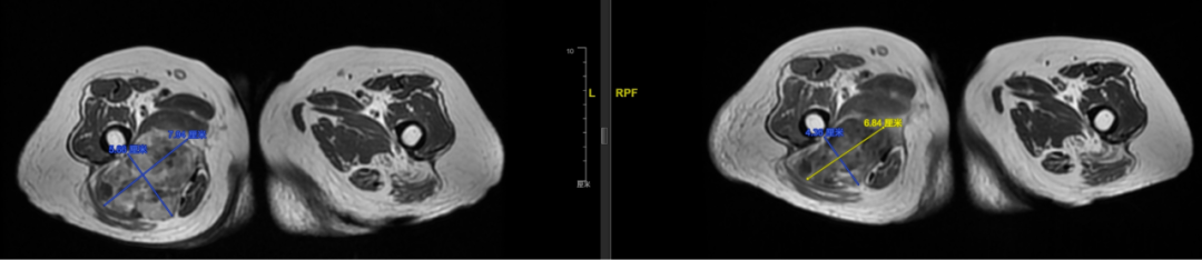
| 2021-12 | MPNST local recurrence | ∼9.0×6.6 cm; surveillance (patient declined intervention) |
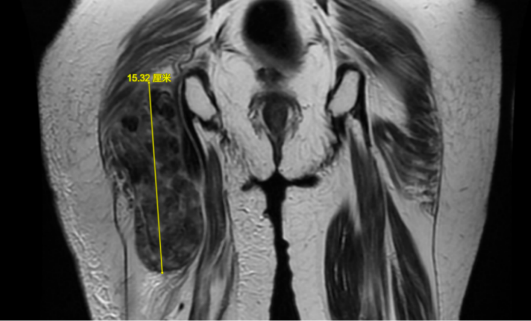
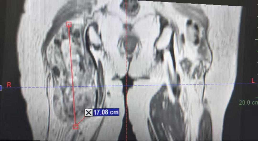
| 2025-06 | MPNST progression with functional impairment | MRI: 8.5×4.8×15.8 cm; bilateral lung mets + pleural effusion |
II. Failed Systemic Therapies: The Toxicity Barrier
Targeted Therapy Attempt — Anlotinib
The patient was started on oral anlotinib, a multi-target tyrosine kinase inhibitor with anti-angiogenic activity used in soft tissue sarcoma.
Outcome: Severe treatment-related adverse events developed:
- Gross hematuria
- Bladder stoma / DJ ureteral stent obstruction
- Right-sided hydronephrosis
Anlotinib was permanently discontinued due to unacceptable urological toxicity.
Immunotherapy Attempt — Tislelizumab
Following anlotinib failure, the patient received tislelizumab, an anti-PD-1 monoclonal antibody.
Outcome: Serious immune-related and infectious complications:
- Persistent high-grade fever
- Left flank pain (renal angle tenderness)
- Urinary tract infection with possible pyelonephritis
- Acute kidney injury (elevated creatinine, impaired renal function)
After supportive management (antipyretics, antibiotics, hydration), symptoms gradually resolved. However, tislelizumab was also discontinued, and the patient explicitly refused any further systemic anti-cancer medications.
III. Imaging Findings and Disease Burden
Baseline CT at Presentation
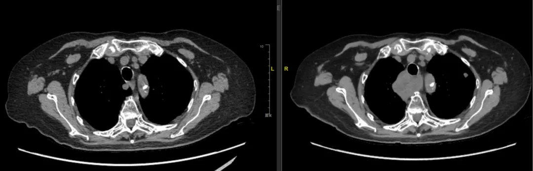
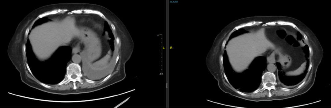
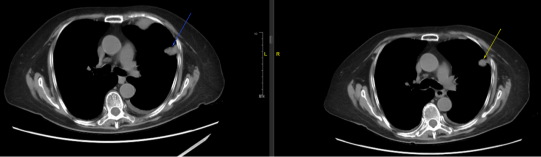
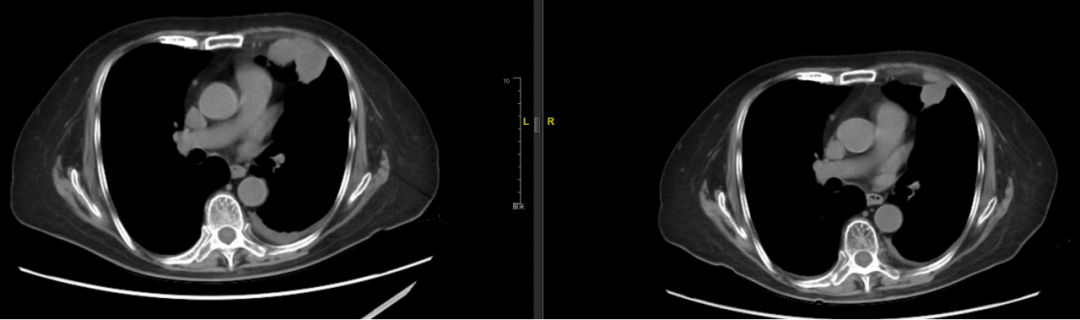
- Lungs: Multiple bilateral pulmonary nodules consistent with metastatic disease. Left chest wall lesions are particularly numerous and bulky.
- Pleura: Small left-sided pleural effusion, likely secondary to left chest wall metastatic involvement.
- General condition: Patient's mental status, sleep, appetite, and weight remained stable — but walking function was severely compromised by the thigh mass.
IV. Core Clinical Dilemmas
Dilemma Matrix
| Challenge Factor | Detail | |
|---|---|---|
| Extreme age | 89 years old; diminished organ reserve; very low tolerance for treatment toxicity | |
| Multiple primaries | Colon cancer, bladder cancer, MPNST — complex overlapping history | |
| Drug intolerance | Both targeted (anlotinib) and immunotherapy (tislelizumab) caused severe AE requiring discontinuation | |
| Local progression | Giant thigh tumor (15.3 cm) causing ambulatory disability requiring urgent symptom relief | |
| Patient preference | Refused biopsy, refused all systemic therapy; accepted only gentle local treatment |
V. MDT Decision: Why Precision Radiotherapy Was Chosen
Treatment Goal Hierarchy
- Control local tumor growth in the right thigh to relieve compressive symptoms
- Restore ambulatory function — enable the patient to walk independently again
- Preserve quality of life above all other endpoints
- Avoid serious adverse events given extreme age and comorbidities
- Address left pleural disease to prevent/treat effusion and preempt future pain from rib/intercostal nerve invasion
Options Evaluated
| Option | Pros | Cons |
|---|---|---|
| Continue systemic therapy (chemo/targeted/immuno) | — | Already proven intolerable; hematuria, hydronephrosis, AKI — life-threatening risks at age 89 |
| Surgical resection | Potential for complete local removal | Tumor >15 cm, deep location, multiple prior surgeries; excessive trauma, high complication risk, poor wound healing in an 89-year-old |
| Best supportive care | No treatment risk | Tumor will continue progressing; complete loss of ambulation imminent; rapid QoL decline |
| Dual-site precision SBRT ✓ SELECTED | Highly localized effect; minimal systemic toxicity; can shrink tumor and relieve compression; aligns with patient's refusal of systemic drugs; simultaneous chest wall coverage may reduce effusion | MPNST has modest intrinsic radiosensitivity; risk of radiation pneumonitis (mitigated by modern planning) |
VI. Radiotherapy Execution: Technical Details
First Course of SBRT (Started December 8, 2025)
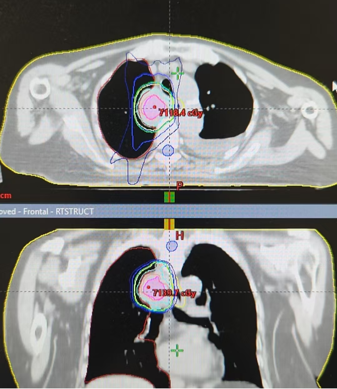
Modality: Stereotactic body radiotherapy (SBRT) — hypofractionated, highly conformal external beam approach.
Site 1: Right Thigh Primary Tumor
| Structure | Definition | Dose |
|---|---|---|
| GTV | Gross tumor volume (visible right thigh mass) | — |
| PGTV | GTV + 5 mm margin | 50 Gy / 10 fractions |
| Boost (GTV – 8mm) | Simultaneous integrated boost to central tumor core | 57 Gy / 10 fractions |
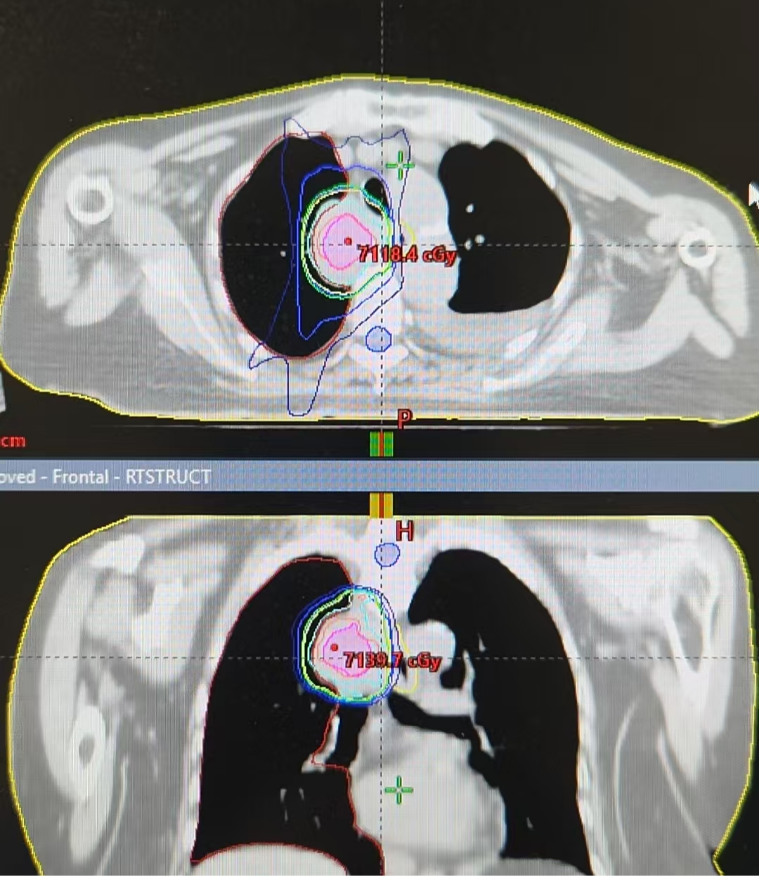
Site 2: Left Pleural Metastasis
| Structure | Definition | Dose |
|---|---|---|
| GTV | Left pleural/chest wall metastatic lesion(s) | — |
| PGTV | GTV + 3 mm margin | 40 Gy / 5 fractions |
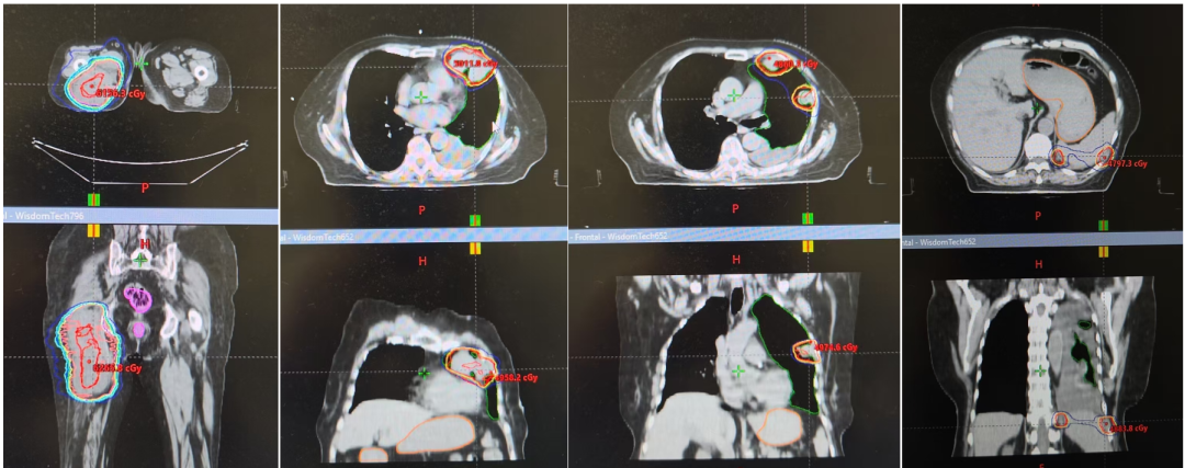
VII. Treatment Tolerance and Toxicity Profile
A critical question for any treatment in an 89-year-old: how well did the patient tolerate it?
| Toxicity Parameter | Result |
|---|---|
| Gastrointestinal | Grade 1 (mild); no Grade ≥ 2 events |
| Hematologic (bone marrow) | No myelosuppression |
| Infectious complications | No infections; no UTI recurrence despite bladder cancer history |
| Cardiopulmonary | No cardiac abnormality; no radiation pneumonitis |
| Constitutional | No fatigue beyond mild |
| Dermatologic (skin reaction) | Grade 1–2 (RTOG criteria) at 1.5 months post-RT; resolved by 3 months (see Figures below) |
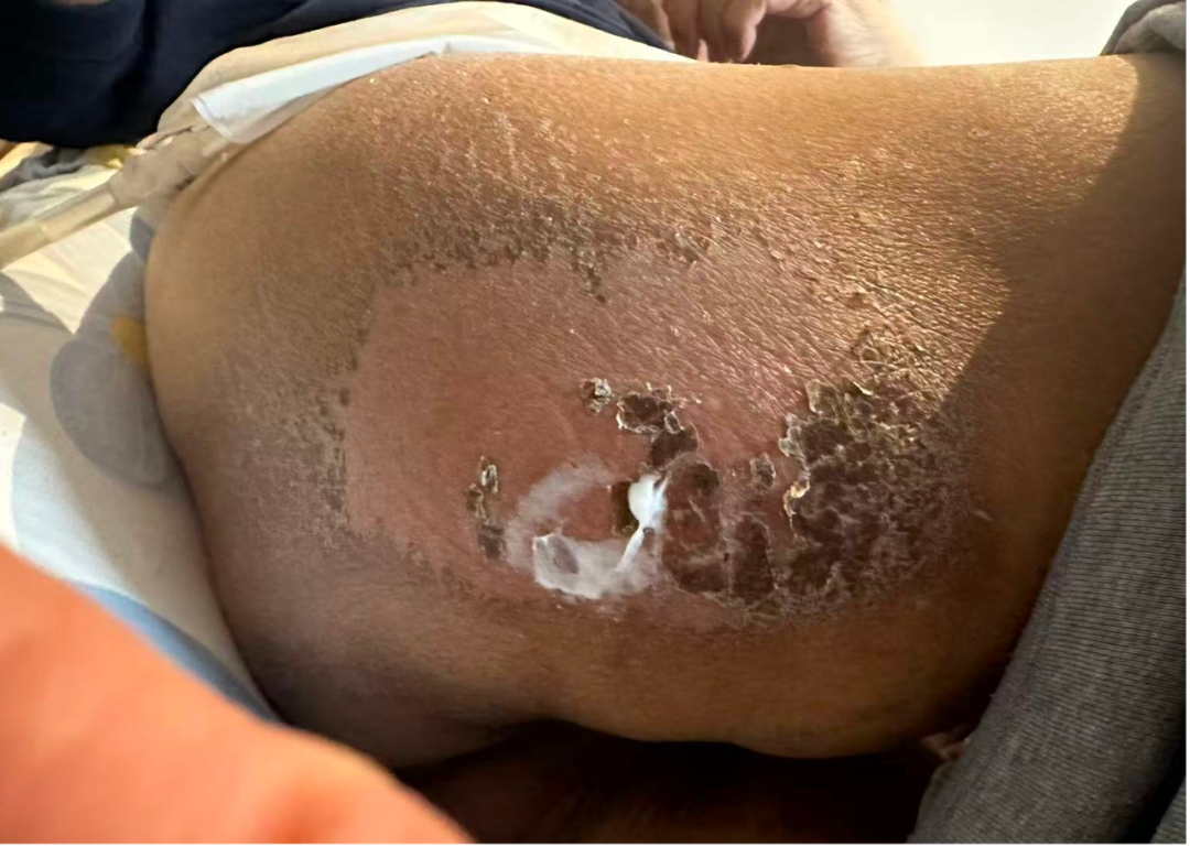
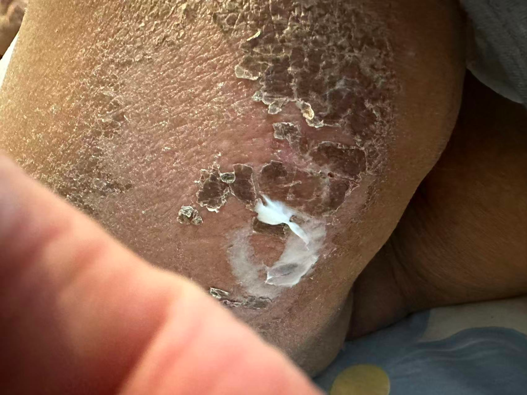
VIII. Clinical Outcomes: Functional Recovery Achieved
Response at 3-Month Follow-Up
- Right thigh tumor: Gradually shrinking since completion of radiotherapy
- Left chest wall lesions: Reduced in size
- Pleural effusion: Decreased compared to baseline
- Symptomatic improvement: Compressive symptoms in the thigh markedly relieved
- Functional outcome: Patient was able to walk independently again within 2 months of completing radiotherapy
- Systemic status: Mental status, appetite, sleep, and body weight all remained stable throughout
Second Course of Radiotherapy (February 2026 Follow-Up)
At the 2-month follow-up chest CT after the first RT course, a previously noted but untreated right pulmonary nodule was found to have rapidly enlarged, now invading toward the mediastinum and approaching the esophagus closely. Although the patient reported no dysphagia at this stage, the trajectory was concerning.
Second-course SBRT plan:
| Structure | Dose |
|---|---|
| PGTV | 52.5 Gy / 15 fractions |
| GTV-integrated boost | 67.5 Gy (simultaneous integrated boost) |
Tolerance of second course: Grade 1 esophagitis during treatment, managed with oral Kangfuxin solution (a Chinese herbal mucosal repair preparation). No other significant symptoms. Patient maintained self-care ability throughout.
IX. Key Clinical Insights
1. Rethinking Cancer Treatment in Extreme Old Age
For patients aged 89 years and older, the clinical mindset must shift away from aggressive cure-oriented strategies toward "functional preservation, symptom relief, and quality-of-life optimization". In this case, abandoning radical intent in favor of precise local control yielded a far better real-world outcome than persisting with poorly tolerated systemic therapies would have.
2. Radiotherapy as the Optimal Salvage Modality
When systemic therapy is exhausted by toxicity and surgery is prohibitive by risk, modern stereotactic body radiotherapy emerges as uniquely positioned to deliver meaningful disease control with minimal systemic burden. Current precision RT technology enables pre-treatment quantitative prediction of organ-at-risk doses, making truly individualized risk-benefit assessment possible before a single fraction is delivered.
3. Managing Multiple Primary Malignancies: Prioritize by Functional Impact
Patients with multiple primary cancers require careful stratification of which disease poses an immediate threat to quality of life versus which may be indolent. In this case, the thigh MPNST — though histologically the most recent diagnosis — was the clear priority because it directly determined whether the patient could walk. The left pleural disease was addressed concurrently because it threatened future complications (effusion, pain). Other smaller pulmonary nodules were monitored rather than overtreated.
4. Patient-Centered Decision Making
This patient refused biopsy, refused systemic drugs, and accepted only "gentle local treatment." Rather than viewing these preferences as obstacles, the treatment team embraced them as binding constraints and designed the optimal solution within those boundaries. Respect for patient autonomy is itself a therapeutic intervention.
5. Soft Tissue Sarcoma: Local RT Has Underappreciated Value
Recurrent, large-volume, high-grade soft tissue sarcomas in elderly patients are often deemed untreatable. This case demonstrates that even for a 15-cm MPNST in an 89-year-old, well-planned local radiotherapy can achieve meaningful tumor debulking, restore lost physical function, and deliver high-quality survival — outcomes that should not be dismissed simply because they fall short of pathological complete response.
Expert Commentary
Prof. Cai Yong
Department of Radiation Oncology, Peking University Cancer Hospital, Beijing
This patient presents as an elderly individual with a history of multiple primary malignant tumors who developed recurrence and metastasis. Based on her medical history and the pattern of recurrence/metastasis, the disease is most likely attributable to the malignant peripheral nerve sheath tumor (MPNST). If surgically feasible, complete resection would be the preferred treatment; however, the efficacy of other systemic therapies for this entity remains limited.
The location of the recurrent and metastatic disease had severely impacted the patient's quality of life. After evaluation, surgical resection was not appropriate, and attempted targeted and immunotherapies produced toxicities that could not be tolerated. At this juncture, a relatively effective yet low-toxicity treatment modality was needed to alleviate the patient's suffering — radiotherapy was unequivocally the best choice.
Modern precision radiotherapy technology allows comprehensive pre-treatment assessment of the probability of various toxicities occurring, enabling precise and safe treatment delivery. The patient's ultimate outcome provides the most compelling validation of this approach.
Importantly, age alone is not an absolute contraindication to cancer treatment. However, given the reduced physiological reserve across organ systems in very elderly patients, we should select modalities that minimize impact on vital organs — and radiotherapy fulfills this requirement exceptionally well. It plays an increasingly important role in the management of geriatric oncology patients.
One consideration for this patient's history of multiple primary malignancies: if feasible, further genetic testing should be pursued to exclude Lynch syndrome (hereditary nonpolyposis colorectal cancer), which would carry implications for family members and potentially inform surveillance strategies for the patient herself.
Seeking Geriatric Oncology or Complex Sarcoma Care in China?
Beijing Arion Cancer Hospital's Radiotherapy Center specializes in stereotactic body radiotherapy (SBRT), image-guided radiotherapy (IGRT), and personalized radiation treatment plans for elderly patients with complex oncologic histories — including multiple primary malignancies, treatment-refractory disease, and cases where systemic therapy options have been exhausted due to toxicity. Our multidisciplinary team integrates radiation oncology, surgical oncology, medical oncology, radiology, and supportive care to design individualized strategies centered on functional preservation and quality of life. International patient services coordinate your complete journey from remote consultation through treatment and long-term follow-up.
Request a Consultation International Patient Guide