
Introduction: A Unique Entity in the Lymphoma Landscape
In the world of lymphomas, Primary Mediastinal Large B-Cell Lymphoma (PMBCL) occupies a truly singular position. Classified under the umbrella of diffuse large B-cell lymphoma (DLBCL) within the non-Hodgkin lymphoma family, it nevertheless exhibits remarkable biological, molecular, and clinical overlap with the nodular sclerosis subtype of classic Hodgkin lymphoma. Medical literature frequently describes it as a disease "standing between non-Hodgkin and Hodgkin lymphomas" — a transitional entity that bridges two major lymphoma classification systems.
Despite its intimidating nomenclature, understanding this disease is essential for patients and clinicians alike because it represents a highly curable malignancy. With accurate diagnosis and standardized therapy, most patients achieve long-term survival — many reaching functional cure.
Case Presentation
Patient Profile
Patient: Female, 45 years old.
Chief Complaint: Left anterior chest wall mass for over one month.
History of Present Illness
The patient developed anterior chest pain approximately one month prior to presentation, followed by the appearance of a progressively enlarging mass on the left anterior chest wall, accompanied by swelling of the left arm. She denied head/neck edema, hoarseness, or dyspnea.
Superficial mass ultrasound: Solid mass in the left anterior-superior mediastinum with malignant features, invading the intercostal muscles of the chest wall, with enlarged lymph nodes adjacent to the thoracic aorta.
PET-CT: Mass-like occupying lesion in the anterior-superior mediastinum with abnormally elevated metabolic activity, initially considered likely of thymic origin. Metastatic lymphadenopathy noted in the right superior mediastinum (paravascular region) and right internal mammary artery region. The lesion showed intimate involvement with surrounding structures, invading the left anterior chest wall and mediastinal vascular spaces, with a small pericardial effusion.
Serum tumor markers: All negative. LDH 244 U/L (↑), α-HBDH 192 U/L (↑).
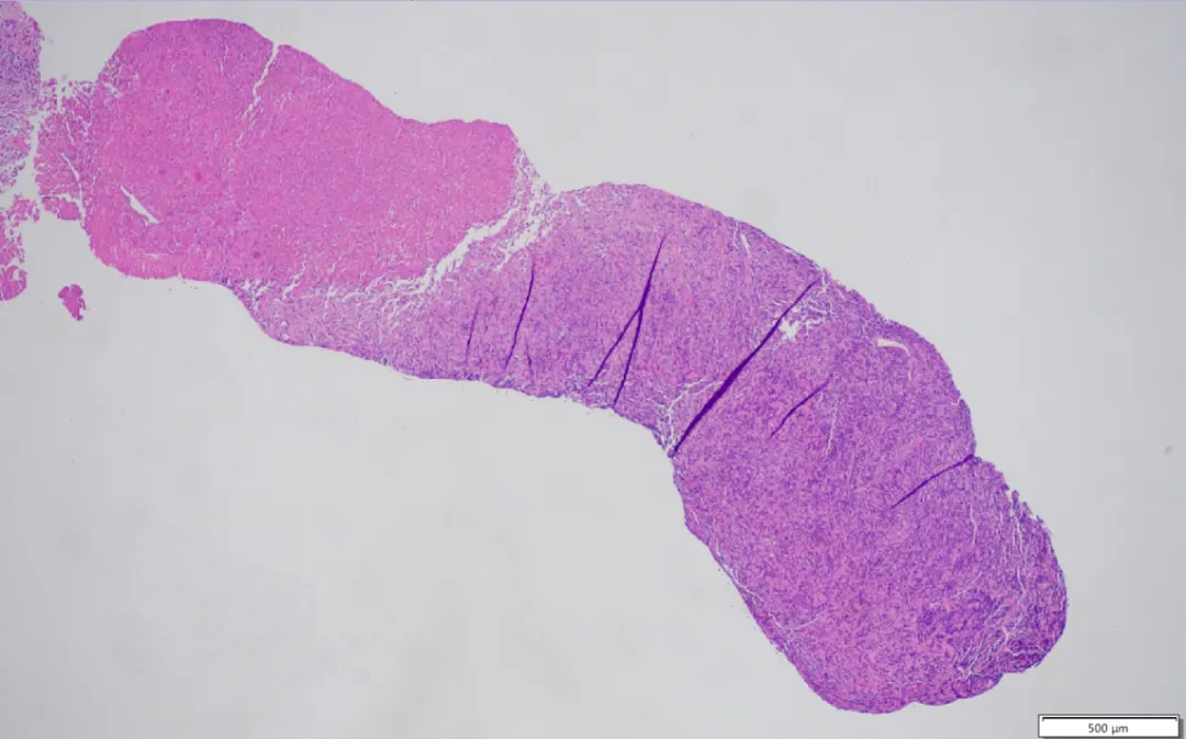
Chest wall core needle biopsy: Microscopy revealed diffusely distributed tumor cells with extensive coagulative necrosis.
At higher magnification, tumor cells were medium-to-large in size, round, with deeply stained nuclei and scant cytoplasm.
Immunohistochemical Profile
Given the wide differential diagnosis for mediastinal masses, comprehensive immunohistochemical (IHC) staining was performed:
| Marker | Result | Significance |
|---|---|---|
| CD20 | Positive | B-cell lineage confirmation |
| MUM1 | Positive | Non-germinal center B-cell phenotype |
| Bcl-6 | Positive | Germinal center marker (variably expressed) |
| Bcl-2 | Positive | Anti-apoptotic protein overexpression |
| Ki67 | Very high proliferation index | Indicates highly aggressive proliferation |
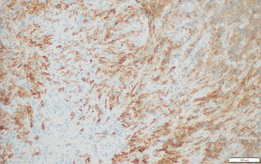
| CD30 | Positive | The hallmark "crossover" marker linking PMBCL to Hodgkin lymphoma |
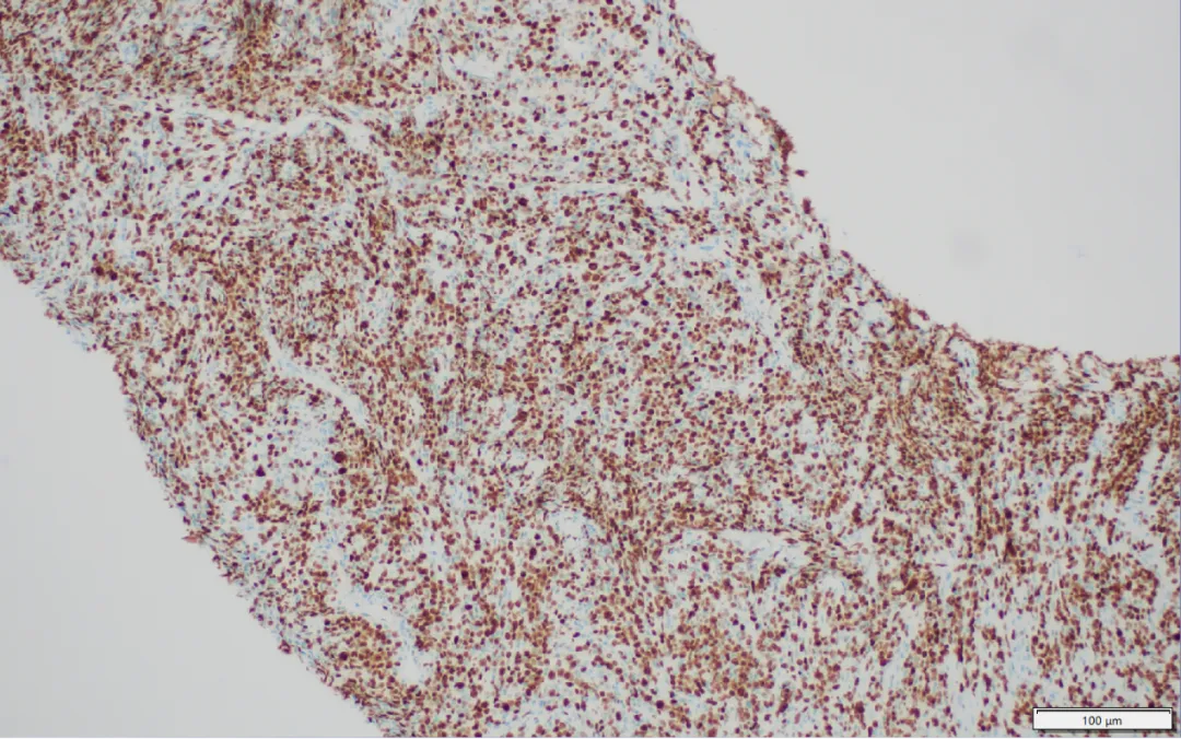
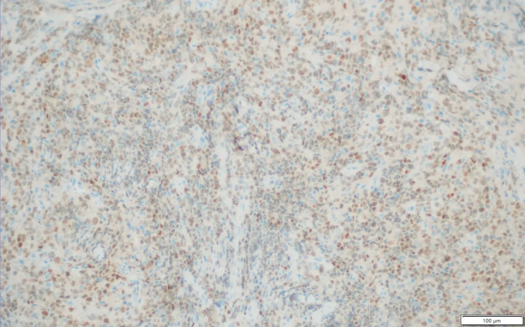
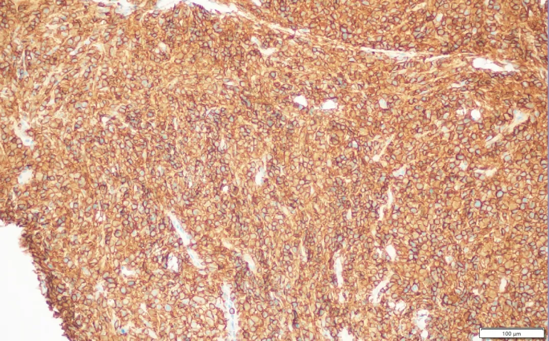
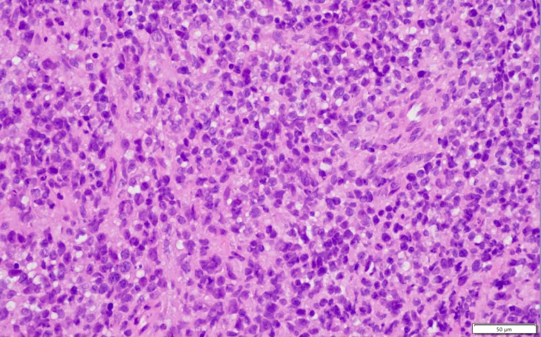
The co-expression pattern of CD20+ (B-cell markers) plus CD30+ (a classic Hodgkin lymphoma-associated marker), set against a fibrotic background rich in inflammatory cells, is the immunophenotypic signature that defines PMBCL as a true biological hybrid. Notably, most cases are CD15-negative and EBV-negative, which helps distinguish PMBCL from classic Hodgkin lymphoma.
I. Where Does It Come From? — Anatomical Origin Shapes Behavior
The name itself tells the story: "Primary" indicates the tumor originates de novo in the mediastinum; "Mediastinum" refers to the compartment between the two lungs, housing the heart and great vessels; "Large B-Cell Lymphoma" denotes its origin from B-lymphocytes with large, actively proliferating cells.
PMBCL is believed to arise from a unique population of thymic B-cells. Although the thymus is principally the organ of T-cell development, it also harbors a small contingent of B cells. These "thymic B-cells" confer upon PMBCL its distinctive biological character, setting it apart from conventional DLBCL arising in lymph nodes or other organs.
This mediastinal origin also explains why PMBCL almost invariably presents as a large anterior mediastinal mass — its most characteristic radiological signature.
II. Who Is Affected? — Young Women Predominate
PMBCL has a striking demographic profile:
- Age of onset: Predominantly 20–40 years old.
- Sex distribution: Females significantly outnumber males.
This stands in sharp contrast to ordinary DLBCL, which typically afflicts middle-aged to elderly individuals. In clinical practice, when a young woman presents with an anterior mediastinal mass accompanied by chest tightness, dyspnea, coughing, or facial/neck edema, PMBCL should rank high on the differential diagnosis list.
III. Clinical Presentation — Not Starting With "Swollen Lymph Nodes"
Unlike many lymphomas that announce themselves through palpable superficial lymphadenopathy, PMBCL's debut is usually a constellation of mediastinal mass compression symptoms:
- Chest tightness, chest pain, shortness of breath
- Persistent dry cough
- Hoarseness (recurrent laryngeal nerve compression)
- Facial or neck swelling (superior vena cava syndrome)
- Palpitations or pericardial effusion
Some patients have no obvious superficial lymphadenopathy and only discover the mediastinal mass incidentally during imaging for breathing difficulties or chest discomfort. A subset may experience constitutional "B symptoms": unexplained fever, night sweats, and significant unintentional weight loss — indicators of biologically active disease.
IV. Imaging Features — An Aggressive Mediastinal Mass
On CT or MRI, PMBCL typically manifests as:
- A large solid mass in the anterior mediastinum
- Irregular morphology; borders may be relatively well-defined or partially ill-defined
- Frequent invasion or encasement of great vessels
- Compression of the trachea, heart, and superior vena cava
- Areas of necrosis or low-density regions within the mass
Imaging can strongly suggest malignancy, but definitive diagnosis requires histopathological examination. Thymoma, germ cell tumors, and metastatic carcinoma may appear similarly on imaging.
V. Why Is It Described As "Between Hodgkin and Non-Hodgkin"?
Under the microscope, PMBCL tumor cells display:
- Large cell size with irregular, prominent nuclei
- Open chromatin with conspicuous nucleoli
- Distinct fibrous septa separating cell clusters
- A background rich in inflammatory cell infiltrate
The immunophenotype reveals a striking hybrid quality: stable expression of canonical B-cell markers (CD20, PAX5, CD79a) combined with weak-to-moderate expression of CD30 — a marker quintessentially associated with classic Hodgkin lymphoma. Most cases lack CD15 expression and are EBV-negative, which distinguishes them from classic Hodgkin lymphoma.
Molecular studies have uncovered shared aberrations between PMBCL and Hodgkin lymphoma:
- 9p24.1 amplification
- PD-L1/PD-L2 overexpression
- JAK-STAT pathway activation
This molecular overlap explains why immune checkpoint inhibitors play a significant role in relapsed/refractory PMBCL.
VI. Diagnostic Challenges — A Master of Disguise
PMBCL's principal mimics include:
- Classic Hodgkin lymphoma (nodular sclerosis subtype)
- DLBCL, other subtypes
- Gray zone lymphoma (true intermediate B-cell lymphoma with features intermediate between DLBCL and Hodgkin)
- Mediastinal thymoma
- Germ cell tumor
Accurate diagnosis demands adequate tissue sampling, integrated interpretation of morphology + immunohistochemistry + molecular testing, and assessment by an experienced hematopathologist. In mediastinal biopsies where tissue is limited, misdiagnosis or missed diagnosis is a real risk.
VII. Treatment Paradigm — Aggressive but Curable
Although classified as a highly aggressive lymphoma, PMBCL is simultaneously among the most curable lymphoma subtypes. Current therapeutic strategies include:
1. Immunochemotherapy as Foundation
Standard regimens include R-CHOP and DA-EPOCH-R (dose-adjusted etoposide, prednisone, vincristine, cyclophosphamide, doxorubicin, rituximab). Many studies consider DA-EPOCH-R a preferred approach for PMBCL, potentially reducing dependence on consolidative radiotherapy.
2. The Diminishing Role of Radiotherapy
Historically, mediastinal radiotherapy was standard after chemotherapy. However, given that most patients are young women — for whom thoracic irradiation increases long-term risks of breast cancer, cardiovascular disease, and lung injury — current practice emphasizes achieving complete remission through intensified chemotherapy before considering whether radiotherapy is necessary.
3. New Hope for Relapsed/Refractory Disease
For patients who relapse or prove refractory, emerging therapies including PD-1 inhibitors (nivolumab, pembrolizumab), CAR-T cell therapy, and targeted anti-CD30 or anti-CD19 agents have significantly improved outcomes.
VIII. Prognosis — Better Than You Might Expect
With guideline-concordant treatment, 5-year overall survival for newly diagnosed patients reaches 80–90% or higher, with many achieving durable, long-term disease-free survival. PMBCL is among the more favorable subtypes of aggressive lymphoma in terms of cure potential.
Prognostic factors include tumor bulk, presence of superior vena cava syndrome, LDH level, and whether complete remission is achieved. In sum, this disease is far less formidable than the word "cancer" might suggest.
IX. Why "Between Two Types of Lymphoma"?
PMBCL defies simple categorization across three dimensions:
- Morphologically: Resembles both DLBCL (large B-cells) and nodular sclerosis Hodgkin lymphoma (fibrous bands, inflammatory background).
- Immunophenotypically: Co-expresses B-cell markers (CD20+) and Hodgkin-associated markers (CD30+).
- Molecularly: Shares 9p24.1 amplification and JAK-STAT activation with Hodgkin lymphoma.
It is not purely non-Hodgkin, nor purely Hodgkin — it is a distinct entity with dual lineage heritage.
X. Take-Home Message
Primary Mediastinal Large B-Cell Lymphoma is a predominantly young-woman's disease originating in the mediastinum, with unique biology, yet it remains highly curable. Its specialness lies not in deadliness but in how precisely modern oncology has come to understand lymphoma subclassification.
It reminds us that cancer is not a monolithic concept but a collection of hundreds of diseases with vastly different "personalities" — and PMBCL is among the most representative: aggressively growing yet carrying tremendous hope; complex to classify yet remarkably optimistic in outcome.
Expert Commentary
Prof. Xue Weicheng
Department of Pathology, Peking University Cancer Hospital, Beijing
Primary Mediastinal Large B-Cell Lymphoma is a B-cell lymphoma with features spanning both Hodgkin and non-Hodgkin classifications, characterized by occurrence in a specific population (young women), at a specific anatomical site (mediastinum), presenting as an exceptionally large mass with firm consistency due to fibrosis, displaying a characteristic immunophenotype (CD30-positive), exhibiting extremely rapid proliferation, and — critically — demonstrating particularly favorable response to treatment and excellent prognosis. Precise pathological diagnosis is of paramount importance.
The immunohistochemical profile observed in this case — CD20+, MUM1+, Bcl-6+, Bcl-2+, high Ki67, and CD30+ — constitutes the textbook signature of PMBCL. The presence of CD30 expression is the key discriminant that links this entity to the Hodgkin spectrum, while the strong B-cell program confirms its placement within the DLBCL family. Recognizing this dual identity is essential for directing appropriate therapy, as PMBCL treatment algorithms differ meaningfully from those for either classic DLBCL or classic Hodgkin lymphoma.
Seeking Advanced Lymphoma Care in China?
Beijing Arion Cancer Hospital offers world-class expertise in diagnosing and treating complex lymphomas including PMBCL, with state-of-the-art pathology, multidisciplinary tumor boards, cutting-edge immunotherapy (including CAR-T and bispecific antibodies), and dedicated international patient services supporting your entire medical journey to China.
Request a Consultation International Patient Guide