Introduction: Common Breast Concerns
Breast-related symptoms are among the most common reasons women seek medical evaluation. Many people discover a lump during self-examination, notice nipple discharge, or receive an abnormal mammogram result and worry that it might be cancer. Understanding what different findings mean — and when they warrant further investigation — can help reduce unnecessary anxiety and guide informed decisions.
This article provides an educational overview of three common breast concerns: breast nodules (lumps), nipple discharge, and the basics of breast cancer screening. It is intended for general health education and is not a substitute for professional medical advice.
Breast Nodules: Benign vs. Concerning Features
A breast nodule is a discrete lump or mass that feels different from the surrounding breast tissue. The vast majority of breast lumps are benign. The key question is: which features suggest a nodule is more likely benign, and which call for prompt evaluation?
- Features often seen in benign nodules: Smooth borders, mobility (easily moves under the skin), pain or tenderness that varies with the menstrual cycle, and stability or shrinkage over time. Common benign conditions include fibroadenomas, cysts, and fibrocystic changes.
- Features that warrant further investigation: Hard or irregular texture, fixation to surrounding tissue, non-tender, progressive enlargement over weeks to months, or associated skin changes such as dimpling or redness.
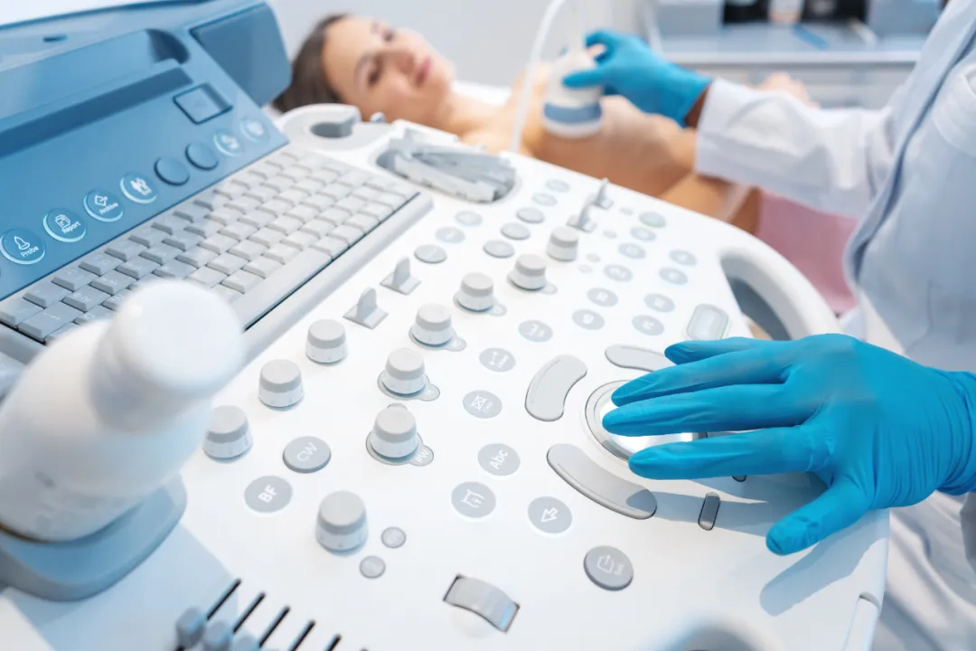
Imaging — typically ultrasound in younger women and mammography in older women — is the first step in characterizing a palpable lump. The BI-RADS (Breast Imaging Reporting and Data System) classification, ranging from 0 to 6, helps standardize the assessment and guides whether a biopsy is needed.
Nipple Discharge: When to Be Concerned
Nipple discharge — fluid leaking from one or both nipples — can be alarming, but not all discharge signals a serious problem. The characteristics of the discharge provide important clues:
- Physiological discharge: Bilateral (both sides), milky or clear, and expressed only with squeezing. This is often related to hormonal fluctuations, medication side effects, or benign duct ectasia.
- Pathological discharge: Spontaneous, persistent, from a single duct on one side, and particularly if the fluid is bloody or serous (watery-straw colored). These features increase the likelihood of an intraductal lesion such as a papilloma or, less commonly, ductal carcinoma.
When pathological discharge is present, evaluation typically includes breast ultrasound, mammography, and in selected cases, breast ductoscopy — a fiberoptic technique that allows direct visualization inside the milk ducts. Ductoscopy can identify and, when appropriate, guide the removal of small intraductal lesions through a minimally invasive approach.
Breast Cancer Screening: An Overview
Screening aims to detect breast cancer before symptoms appear, when treatment is most likely to be effective. The main screening tool is mammography, supplemented by ultrasound in women with dense breast tissue.
General screening recommendations include:
- Average-risk women: Annual or biennial mammography starting at age 40–50, depending on national guidelines and individual risk factors. Clinical breast examination may complement imaging.
- Higher-risk women: Those with a strong family history of breast or ovarian cancer, known BRCA1/BRCA2 mutations, or prior chest radiation may begin screening earlier (age 25–30) and may benefit from breast MRI in addition to mammography.
If a screening mammogram shows an abnormality, further workup — diagnostic mammography with spot compression views, targeted ultrasound, and when indicated, core needle biopsy — is performed to determine whether the finding is benign or malignant.

Minimally Invasive Diagnostic Techniques
Advances in breast diagnostics have introduced tools that can obtain tissue diagnosis with less discomfort and better cosmetic outcomes than traditional open surgical biopsy:
- Core needle biopsy (CNB): Uses a hollow needle to extract small cylinders of tissue from a suspicious area, typically under ultrasound or stereotactic guidance.
- Vacuum-assisted biopsy (VAB): A device that uses suction to obtain a larger tissue sample through a single needle insertion, improving diagnostic accuracy for microcalcifications and small lesions.
- Breast ductoscopy: As mentioned above, this technique is used for evaluating pathological nipple discharge by directly visualizing the ductal system and obtaining targeted biopsies of intraductal lesions.
These techniques reduce the need for open surgical biopsy in many cases, while providing sufficient tissue for definitive pathological diagnosis including hormone receptor status (ER/PR) and HER2 testing when cancer is found.
Key Points to Remember
- Most breast lumps are benign. Discovering a lump does not mean you have cancer, but it does mean you should have it evaluated by a healthcare provider.
- Know what is normal for you. Breast self-awareness — understanding how your breasts normally look and feel — helps you notice changes early.
- Nipple discharge characteristics matter. Spontaneous, unilateral, bloody, or serous discharge should be evaluated promptly.
- Screening saves lives. Regular mammography according to age- and risk-appropriate guidelines is the most effective tool for early detection of breast cancer.
- Minimally invasive techniques are widely available. Needle biopsies and ductoscopy can often provide a diagnosis without the need for open surgery.
Medical Disclaimer
This article is provided for general medical education and public health information only. It does not constitute medical advice, diagnosis, or treatment recommendations. Every patient's situation is different; breast health concerns should always be discussed with a qualified physician. If you notice a new breast lump, nipple discharge, or other breast change, please consult a licensed healthcare professional for individualized evaluation.