This article is a de-identified educational case review. It describes the clinical course of one patient managed at Beijing Arion Cancer Hospital and reflects the team's approach to multidisciplinary decision-making around contrast-agent selection and imaging protocol design. It is not medical advice, nor does it guarantee outcomes for other patients. Treatment decisions must be individualized by qualified physicians.
Case Introduction
This issue reviews how a gynecologic oncology patient with suspected pulmonary embolism — who urgently needed pulmonary artery CT angiography (CTA) to confirm or exclude a clot — was managed when a documented iodine contrast allergy ruled out the standard examination. With no clearly superior alternative imaging option available, the team faced a genuinely high-risk dilemma: how to sidestep the iodine contrast contraindication while still completing the CTA quickly enough to answer the clinical question, balancing technical risk against diagnostic safety. Through multidisciplinary (MDT) deliberation, a gadolinium-based contrast agent combined with a "dual-low" scanning protocol and deep-learning reconstruction broke the deadlock.
I. Case Overview: Baseline Status and Core Challenges
1. Baseline Status
Presentation: A 52-year-old woman was admitted with "stage III tubal cancer (poorly differentiated adenocarcinoma), recurrence after postoperative multi-line therapy". She showed multiple abnormal laboratory indices after cancer treatment and a frail general condition, in a hypercoagulable state, with laboratory findings suggesting a possible thrombotic risk — so pulmonary embolism had to be excluded.
Core challenge: An urgent "pulmonary artery CTA" was requested, but the patient had a documented iodine contrast allergy.
2. Current Dilemma
Pulmonary artery CTA is the first-choice examination for diagnosing pulmonary embolism: it conventionally uses an iodine-based positive contrast agent, and the presence or absence of a "filling defect" within the pulmonary artery lumen is used to judge whether embolism is present. It is convenient, rapid and easy to interpret — the preferred examination for clarifying pulmonary artery embolism. However, the patient's documented iodine contrast allergy is a primary contraindication, sharply restricting that examination. Non-iodinated contrast agents have no clear clinical-practice-guideline support, so how to reach the diagnostic goal quickly under controllable risk became the central question on which confirmation of the diagnosis depended.
3. Key Assessment Findings
One year earlier the patient had undergone a contrast-enhanced pelvic MRI, and the examination report confirmed it; the gadolinium contrast agent used then had caused no allergic or other adverse reaction. Today's serum creatinine, converted to an estimated glomerular filtration rate (eGFR), was about 96 mL/min/1.73 m² — within the normal renal-function range. It was therefore considered relatively safe to use the gadolinium contrast agent routinely employed in contrast-enhanced MRI, in place of iodine contrast, to complete this CTPA examination.
II. Decision-Making: MDT Core Analysis and Strategy Deliberation
1. Composition of the MDT Team
Gynecologic Oncology (Internal Medicine); and the Radiology Imaging Centre — including its physician group, technologist group and nursing group.
2. Pathway Deliberation and Trade-off Analysis
Radiology physicians: If pulmonary artery CTA cannot be performed because of allergic risk, could another examination be considered as a substitute?
Gynecologic Oncology: Pulmonary angiography is the gold standard, but it is invasive and costly; could radionuclide ventilation/perfusion scanning (V/Q imaging) or magnetic resonance pulmonary angiography (MRPA) be considered instead?
Radiology physicians: Radionuclide V/Q scanning requires the radionuclide to be ordered in advance and cannot meet the requirements of an emergency examination.
Radiology technologists: MRPA demands a high level of patient cooperation. This patient is frail; if cooperation is poor, image quality is unstable, and the scan is long with many influencing factors.
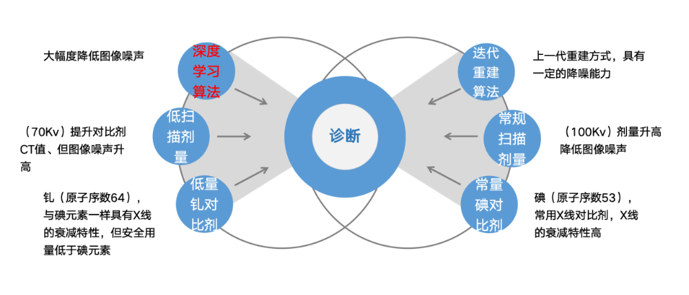
Radiology technologists: The core imaging element of the gadolinium contrast agent used in contrast-enhanced MRI is gadolinium. Gadolinium and iodine are both high-atomic-number elements; according to the characteristics of the X-ray absorption curve for high-atomic-number elements, gadolinium contrast can in principle be used as a positive contrast agent for X-ray examinations. However, because gadolinium's X-ray absorption efficiency is lower than iodine's, its contrast efficacy as an X-ray positive contrast agent is markedly weaker, and the safe dose of gadolinium contrast is markedly lower than that of iodine contrast. Choosing gadolinium in place of iodine therefore faces the challenges of a limited dose and a markedly weakened contrast effect; and adopting a low-kV, low-contrast-dose "dual-low" technique would bring declining image quality and rising image noise. But how low should the "dual-low" parameters be — low enough to be safe and to form good contrast, yet not so low that excessive noise degrades diagnostic detail? A conventional iodine-based pulmonary CTA performed with standard parameters (120 kVp, about 40 mL of iodine contrast) yields a pulmonary artery luminal CT value above 300 HU. This "dual-low" technique would use 20 mL of gadolinium contrast at 80 kVp or lower, and the resulting CT value would need to be at least above 150 HU to form a clear contrast effect on the images. Given that deep-learning technology in new-generation multi-detector spiral CT happens to reduce noise and improve image quality, the examination is technically feasible using gadolinium contrast for pulmonary CTA.
Radiology physicians: Pulmonary artery CTA is the preferred examination for clarifying whether pulmonary embolism is present, but will the image noise after the "dual-low" plus deep-learning technique affect the display of lobar- and segmental-level vessel branches? To improve contrast, could an even lower kVp be attempted? In addition, experienced radiologists should perform a two-reader interpretation to ensure reliability.
Radiology technologists: A 70 kVp parameter can be used, and the gadolinium contrast should be injected at the high flow rate used for iodine contrast to ensure the contrast effect.
Radiology nursing: Using a high-pressure injector with a high-flow indwelling needle together with the examination technique is mature practice; strengthen fixation, avoid extravasation, and monitor the injection pressure curve to ensure the examination proceeds smoothly.
3. Breakthrough Points
The application of CT dual-low scanning technology combined with deep-learning technology was the technical key that turned "feasible" into "possible".
The teamwork of the radiology imaging centre's physicians, technologists and nurses was an important factor in ensuring the examination proceeded smoothly.
III. The Breakthrough: Examination Execution and Technical Points
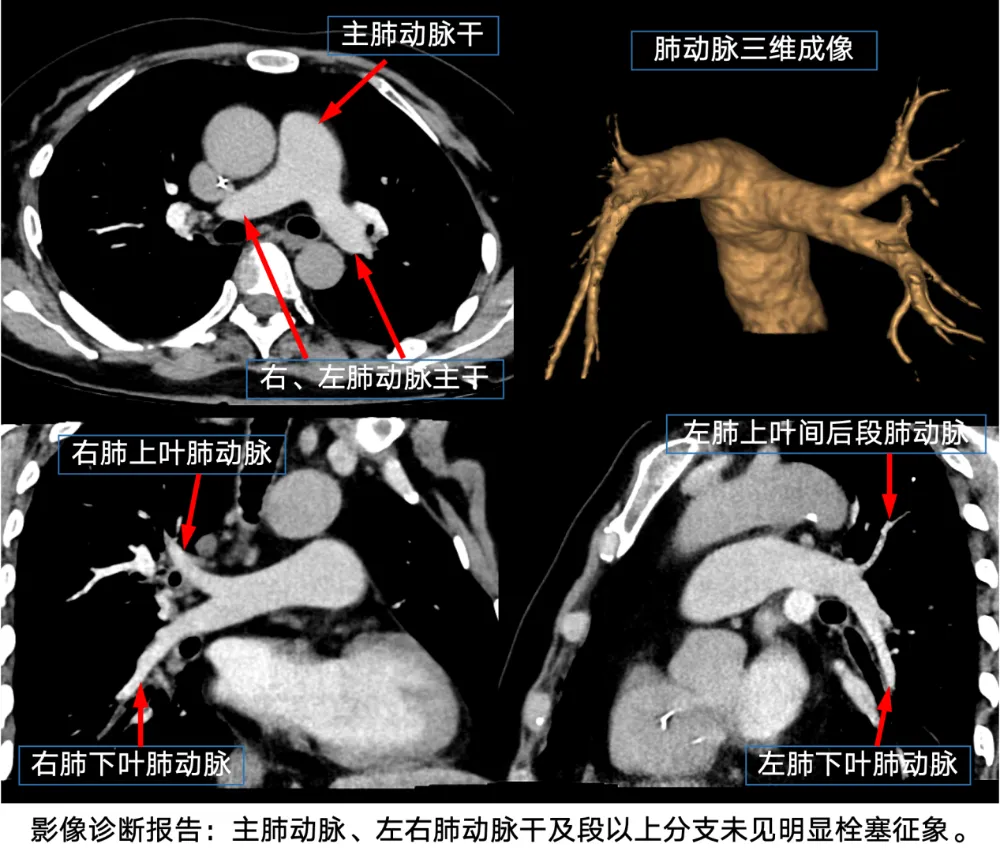
The final examination was performed on a GE Apex 256-row spectral CT using a dual-low scanning technique (70 kVp, 20 mL gadolinium contrast — gadopentetate dimeglumine, gadolinium content 9.38 g), with an injection rate of 5 mL/s and a gantry rotation of 0.28 s, using an axial scan triggered by monitoring at the pulmonary artery origin (trigger threshold 100 HU). After deep-learning algorithm post-processing, the CT values of the pulmonary artery trunk and the lobar pulmonary artery branches were all above 150 HU. Although the sub-segmental and sub-sub-segmental pulmonary artery branches could not be accurately assessed because of contrast-agent distribution and declining image quality, the images were sufficient to meet the clinical diagnostic and treatment needs.
IV. Outcome Assessment
The gadolinium "dual-low" pulmonary CTA was completed safely and yielded an interpretable study: the imaging report confirmed no obvious embolic signs in the main pulmonary artery, the right and left pulmonary artery trunks, or their branches at segmental level and above. The examination answered the urgent clinical question — helping to exclude pulmonary embolism — without exposing the iodine-allergic patient to iodine contrast.
V. Insights from This Case
Through deep excavation of imaging principles, a previously limited dual-low scanning technique was reborn under the combined support of new technology and deep-learning algorithms; coupled with the teamwork of the MDT model, it enabled an individualized examination design for a patient with special conditions, elevating the meaning of the examination itself and creating the conditions for innovative thinking combined with multi-dimensional technology application — actively adapting to clinical diagnostic and treatment needs so that the patient benefits.
Yet the exploratory frontier of technology never ends. Could a spectral imaging approach generate the highest-contrast image at the optimal keV while also providing spectral parameters that offer more diagnostic information — and can image noise be effectively controlled? Could a perfusion scanning approach, using very little contrast, capture the optimal images within the narrowest contrast-peak time window to achieve the best vascular contrast — and can the scanning dose be well controlled? Could a sustainably optimizable algorithm model be built that generates a personalized optimal scanning protocol from the patient's body habitus and vascular-access conditions, the basic parameters of right-heart function and the performance of the CT equipment, with image quality optimized by the algorithm to achieve a perfect image across all metrics? All of these are worth further thought prompted by this case. Refined imaging, striving for perfection — the Radiology Imaging Centre of Beijing Arion Cancer Hospital has always worked toward this.
Expert Commentary
Prof. Feng Feng
Director and Chief Physician of the Department of Radiology, Peking Union Medical College Hospital, Chinese Academy of Medical Sciences; MD, doctoral/postdoctoral supervisor; Chair of the Department of Imaging Medicine and Nuclear Medicine.
This patient faced a pain point frequently encountered in clinical practice. For patients who need contrast-enhanced CT, a history of iodine contrast allergy usually becomes a contraindication to enhancement; if such patients have no good alternative imaging method, they cannot obtain a timely and accurate diagnosis.
For patients with suspected pulmonary embolism, the clinical presentation is often chest pain in the emergency department, requiring prompt clarification of whether a larger-calibre pulmonary artery embolism is present. In such situations other imaging methods cannot readily substitute for the timely, definitive imaging diagnosis that is urgently needed. CT, with its speed and high spatial resolution, has always played an important role in the emergency setting.
This patient was a cancer patient with recurrence after multiple lines of treatment, in poor general condition and a hypercoagulable state, with laboratory findings suggesting possible thrombotic risk, requiring pulmonary embolism to be excluded. But the patient was allergic to iodine contrast. Clinicians also proposed alternative nuclear medicine examinations or contrast-enhanced magnetic resonance pulmonary angiography; however, the radionuclide must be prepared in advance, and MRI takes a long time, making it hard for a frail patient to cooperate well enough to obtain diagnostic-quality images. So taking a different path — using an MRI contrast agent to perform a CTA examination — is indeed an approach worth trying.
According to physical principles, the atomic number of iodine is 53 and that of gadolinium is 64, and their X-ray absorption characteristics differ: the K-edge transition energy is about 33.2 keV for iodine and about 50.2 keV for gadolinium. Conventional CT X-rays have photon energies mostly of 20–80 keV, so both can enhance contrast, and energy CT imaging is also possible. It is just that for gadolinium, photoelectric absorption rises significantly to improve contrast only when photon energy exceeds 50.2 keV. So iodine is still conventionally used as the CT enhancement contrast agent. In addition, gadolinium, as a heavy metal, usually exists in the Earth's crust in inorganic form and is relatively hard to obtain. Furthermore, like iodine contrast agents, different gadolinium contrast agents have different gadolinium concentrations, and their enhancement effects should also differ; further application experience is still needed to refine this examination.
The earliest literature report of using gadolinium contrast for X-ray enhancement examinations was published in the European Journal of Radiology in 1997, applying gadolinium contrast to CTA of the aorta, renal arteries and pulmonary arteries. A more recently published (2026) article reported the feasibility of using gadolinium contrast for pulmonary artery CTA on photon-counting CT. In addition, there have been successful overseas cases of using gadolinium contrast for superselective arterial interventional therapy in patients with severe iodine contrast allergy. However, no such report has yet appeared in the domestic literature.
This patient's attempt to successfully perform a pulmonary artery CTA with gadolinium contrast involved a thorough preoperative MDT discussion; given the patient's frail condition, a dual-low examination mode was chosen to reduce the contrast dose and minimize the radiation dose as far as possible, ultimately achieving a relatively ideal examination result. This has laid a solid foundation for the future use of gadolinium contrast pulmonary artery CTA in patients allergic to iodine contrast.
Medical Disclaimer
This article is provided for general medical education and public health information only. It does not constitute medical advice, diagnosis, or treatment recommendations. Clinical outcomes depend on individual circumstances, and treatment decisions should always be made with a qualified physician.