
This article is a de-identified educational case review. It describes the clinical course of one patient managed at Beijing Arion Cancer Hospital and reflects the team's approach to multidisciplinary decision-making when a rare opportunistic pathogen is suspected. It is not medical advice, nor does it guarantee outcomes for other patients. Treatment decisions must be individualized by qualified physicians.
Case Introduction
This issue presents a case in which a rare pathogen was detected. The patient was diagnosed in June 2025 with squamous cell carcinoma of the right main bronchus, and after concurrent chemoradiotherapy he continued on immune maintenance therapy. In November 2025 his interstitial lung disease (ILD) worsened, and radiation pneumonitis and immune-related pneumonitis could not be excluded, so he was treated with methylprednisolone and his cough and sputum improved. On admission on January 5, 2026, imaging showed a new consolidation in the right upper lobe, a nodular lesion in the left lower lobe and right-sided pleural effusion. On January 9 his condition changed and he coughed up black and yellow sputum. A sputum smear under the microscope revealed Gram-positive branching bacilli; after discussion with the attending physician a deep sputum specimen was re-collected, and Nocardia otitidiscaviarum was successfully cultured. This organism is rare, grows slowly, has a low culture-positive rate in sputum, is highly pathogenic and carries a high mortality — which is precisely why the way it was caught deserves a closer look.
I. Case Overview: Baseline Status and Core Challenges
1. Baseline Status
The patient was a 72-year-old man with a history of old cerebral infarction. In June 2025 he was diagnosed at our hospital with moderately differentiated squamous cell carcinoma of the right main bronchus, staged cT4N2M0, stage III. Because the tumour lay close to the carina, surgery could not achieve a clean margin, so he received concurrent chemoradiotherapy.
He was admitted for the fourth time on November 20, 2025. Chest CT showed that bilateral ILD had worsened markedly compared with before, and some tumour markers were elevated. Radiation pneumonitis was considered, with concomitant immune-related pneumonitis not excluded, so he was given methylprednisolone 80 mg/day together with moxifloxacin for infection prophylaxis. A repeat chest CT on December 4 showed that the ILD had improved, and he was discharged on regular oral methylprednisolone.
On his fifth admission, on January 5, 2026, he developed fever with a peak temperature of 38.6 °C. Laboratory tests showed a white blood cell (WBC) count of 9.98×10⁹/L (elevated), a neutrophil percentage of 92.4% (elevated) and a C-reactive protein (CRP) of 114.3 mg/L (elevated). Imaging showed a new consolidation in the right upper lobe, a nodular lesion in the left lower lobe and right-sided pleural effusion. Infection was considered, so he received anti-infective and expectorant therapy along with a full pathogen workup. Based on susceptibility results, a drug with anti-pseudomonal activity was selected, while coverage for Nocardia was also taken into account. On January 9 his condition changed and he coughed up black and yellow sputum, which was sent for repeat sputum culture.
2. Core Challenges
(1) A rare, dangerous opportunist. Nocardia otitidiscaviarum belongs to the genus Nocardia. Lung infection caused by this genus is called pulmonary nocardiosis, a serious opportunistic infection that occurs mostly in patients with underlying disease and impaired immune function. It can present as pneumonia or lung abscess, and readily spreads haematogenously to other organs — especially the central nervous system. Early diagnosis and early treatment are the key to a good prognosis.
(2) A pathogen that hides from the culture plate. Because Nocardia grows more slowly than most bacteria in culture media, when a sputum specimen is of poor quality the normal upper respiratory flora overgrows and suppresses it, so the culture-positive rate is low. A negative culture means no organism for identification or susceptibility testing — and therefore no early diagnosis to guide clinical treatment.
II. Key Steps in Infection Diagnosis
Microbiological Diagnostic Milestone 1: Read the Smear Carefully — Recognise the Organism
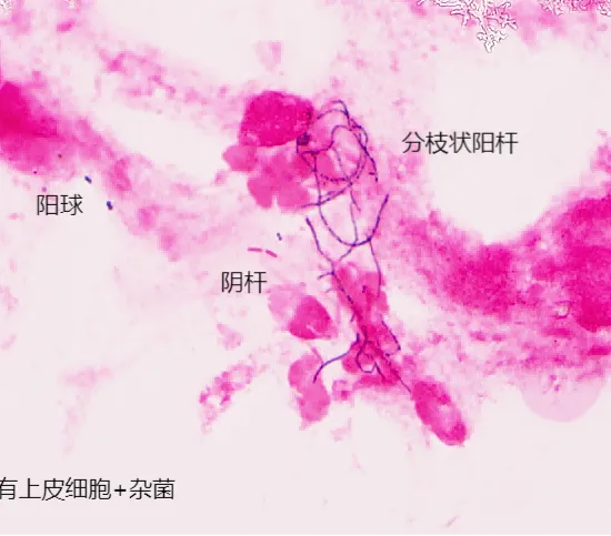
January 9, 2026: the sputum smear under the microscope showed Gram-positive slender branching bacilli, together with Gram-positive cocci and Gram-negative bacilli. Nocardia or nontuberculous mycobacteria (NTM) was suspected, so acid-fast staining and modified acid-fast staining were added for preliminary diagnosis. Because the sputum specimen was of poor quality, however, the normal upper respiratory flora overgrew and the culture was negative.
Microbiological Diagnostic Milestone 2: Communicate Closely to Move Diagnosis Forward
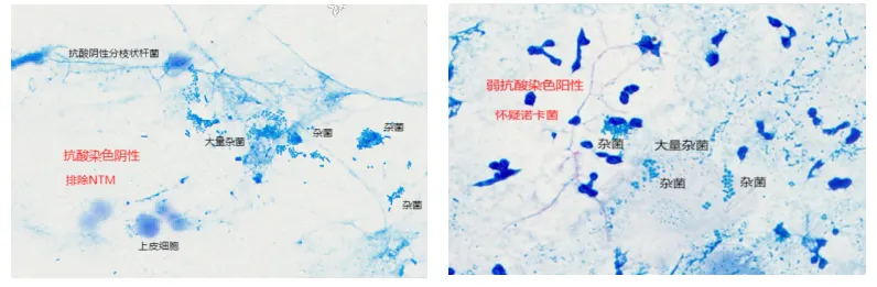
The attending physician discussed the patient's clinical situation with the laboratory, along with the laboratory's progress and its difficulty: the appearance on Gram stain of the sputum smear and the laboratory's suspicion. Subsequently modified acid-fast staining was positive (suspected Nocardia) while acid-fast staining was negative (excluding NTM). After culture, only normal flora was seen — it was presumed that overgrowth of normal flora in a specimen of upper respiratory origin had suppressed the growth of Nocardia.
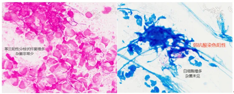
The clinical team took this seriously. On January 12, following the laboratory's recommendation, the patient gargled thoroughly and a deep sputum specimen was re-collected for repeat testing. On microscopy the repeat deep sputum specimen was of good quality: category A sputum, with more than 25 white blood cells per low-power field and fewer than 10 epithelial cells per low-power field; the Gram-positive slender branching bacilli were markedly increased compared with before, and modified acid-fast staining was positive. The laboratory inoculated duplicate media and placed them in a carbon dioxide incubator and an ordinary incubator respectively, because Nocardia grows better in air than in a carbon dioxide environment.
Microbiological Diagnostic Milestone 3: Unravel the Threads — At Last, the Result
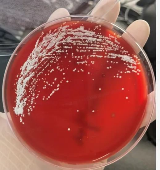
Culture: after 48 hours in the ordinary incubator, the blood agar plate showed white, dry, downy aerial colonies that could grow embedded within the medium itself.
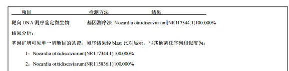
Species identification: because Nocardia is a rare organism, first-generation (Sanger) sequencing was expedited, and susceptibility testing was performed in parallel according to the CLSI M45 requirements for Nocardia. Identification and susceptibility testing were completed together, and the results were reported back to the clinical team promptly. Metagenomic next-generation sequencing (mNGS) of the sputum also confirmed Nocardia otitidiscaviarum.
III. Analysis of the Diagnostic and Treatment Process
1. A high-risk host: the patient's relatively low immunity, his underlying lung disease and his steroid treatment together placed him in the high-risk group for Nocardia infection.
2. Imaging support: imaging clarified the nature of the pulmonary disease progression and supported the presence of new inflammation.
3. Laboratory tests: infection markers were elevated, while tumour markers continued to fall compared with before.
4. Microbiological examination: the sputum smear raised suspicion of the rare organism Nocardia; a repeat high-quality specimen successfully cultured Nocardia; the organism was actively identified to species level and susceptibility testing was completed, providing the clinical team with a basis for anti-infective drug selection.
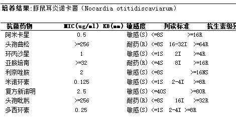
5. Treatment: the susceptibility report showed that this Nocardia otitidiscaviarum isolate was susceptible to trimethoprim-sulfamethoxazole (co-trimoxazole), ciprofloxacin, amikacin, linezolid, minocycline and doxycycline, and resistant to ceftriaxone, imipenem and cefepime. Because co-trimoxazole is also the first choice for Pneumocystis jirovecii, the team used co-trimoxazole combined with moxifloxacin for anti-bacterial treatment.
IV. Outcome Assessment and Follow-up Strategy
1. Short-Term Outcome
The patient's symptoms continued to improve. Chest CT showed that the infectious lesions in the right upper lobe and left lower lobe had shrunk compared with before, and the bilateral interstitial inflammation had been absorbed — indicating that the current treatment was effective. Repeat inflammatory markers PCT, CRP and SAA were all normal. He is currently on methylprednisolone 40 mg/day, with a plan to taper the dose.
2. Subsequent Treatment and Follow-up Plan
After conventional treatment of pulmonary nocardiosis, low-dose, long-course maintenance therapy is very important, because otherwise relapse is very likely. For pulmonary nocardiosis in immunocompromised patients, a course of at least 6 months is recommended.
Follow-up requires periodic chest CT to observe changes in the lesions — which is consistent with the periodic chest CT already needed to monitor this patient's right main bronchus squamous cell carcinoma.
V. Insights from This Case
1. Direct smear microscopy is crucial. In this case the laboratory technician suspected Nocardia after seeing "branching filaments" on the sputum smear, and modified acid-fast staining was then able to reveal it. It is therefore essential to perform an original direct smear examination on all kinds of specimens — it often yields unexpected findings. This also reminds microbiology staff to "recognise the organism with a keen eye", reading smears carefully during staining and never letting a suspicious morphology go.
2. Close clinician–laboratory communication moves diagnosis forward. In this case the laboratory promptly communicated its suspicion to the clinical team and asked for a repeat high-quality specimen; after the repeat deep sputum sample, the target organism was successfully cultured. This also suggests that when clinicians suspect a rare organism, they should actively communicate with the laboratory and ask it to take measures to improve the detection rate — for example, prolonging culture time, or using a suitable medium, or using a suitable incubator.
3. A high-quality specimen is decisive for the test result. Nocardia otitidiscaviarum grows slowly; at first it was masked by relatively fast-growing normal flora or other pathogens and so was missed. Specimen collection must therefore be done properly.
4. Patients with malignant tumours are a high-risk group. After radiotherapy, chemotherapy and immunotherapy — and especially after steroid treatment — a cancer patient's immune function is further impaired, making them a high-risk group for pulmonary nocardiosis. Clinicians should consider the possibility of Nocardia infection in such suspicious cases.
5. Metagenomic sequencing is a good supplementary diagnostic method for rare organisms, but it cannot provide a susceptibility report; culturing live organisms and performing susceptibility testing remains essential for treatment.
6. Pulmonary nocardiosis is easily missed or misdiagnosed. Because its clinical presentation is non-specific and its incidence is low, and because Nocardia grows slowly and is difficult to culture, pulmonary nocardiosis is prone to missed diagnosis and misdiagnosis. The disease has a high mortality and relapse rate, and in patients with respiratory malignant tumours the mortality is even higher. Early diagnosis and effective treatment are therefore the key to improving the prognosis.
Expert Commentary
Prof. Yang Duo
PhD in Clinical Laboratory Diagnostics, Peking University Health Science Center; Chief Technologist.
Nocardia, and especially rare species such as Nocardia otitidiscaviarum, have a low incidence of infection. At the same time such infections occur mostly in immunocompromised people, in whom the clinical presentation often lacks specificity and is easily confused with the underlying disease, while imaging findings are non-characteristic — so the final diagnosis depends on pathogenetic evidence.
Nocardia grows far more slowly than ordinary pathogens. If the specimen is of poor quality (such as the first non-deep sputum sample in this case), the normal upper respiratory flora multiplies rapidly and suppresses its growth, resulting in a low culture-positive rate; and because its morphology resembles that of nontuberculous mycobacteria, routine staining cannot distinguish them quickly — further increasing the difficulty of early pathogenetic confirmation.
In this patient's sputum testing, the first sputum culture showed Gram-positive slender branching bacilli on Gram staining; the possibility of Nocardia or NTM came to mind at once, and acid-fast and modified acid-fast staining were proactively added for differential diagnosis, providing a key clue for the subsequent direction. This reflects the meticulous, professional spirit of not letting any "trace" go in laboratory work. At the same time, the laboratory proactively communicated with the clinical team and pushed for a repeat high-quality clinical specimen — showing that even today, when laboratory technology is highly developed, collaboration between clinicians and the laboratory still plays an important role. For the detection of rare organisms, using metagenomic sequencing for confirmation is a further good example of modern genetic testing technology applied to microbiological examination.
In summary, the successful detection of this rare infection depended on the professional attentiveness of the laboratory staff, good clinical–laboratory collaboration, and the wide application of genetic testing technology. This provides a reliable pathway for the future clinical diagnosis of such difficult and rare infections.
Medical Disclaimer
This article is provided for general medical education and public health information only. It does not constitute medical advice, diagnosis, or treatment recommendations. Clinical outcomes depend on individual circumstances, and treatment decisions should always be made with a qualified physician.