
This article is a de-identified educational case review. It describes the clinical course of one patient managed at Beijing Arion Cancer Hospital and reflects the team's approach to multidisciplinary decision-making across the disease trajectory. It is not medical advice, nor does it guarantee outcomes for other patients. Treatment decisions must be individualized by qualified physicians.
Case Introduction
Breast neuroendocrine carcinoma (NECB) is a rare breast malignancy, and among these the high-grade small-cell neuroendocrine carcinoma (SCNEC) is particularly aggressive, with clinical management still facing major challenges. This issue focuses on a 42-year-old woman diagnosed at the outset with mixed high-grade SCNEC of the breast. Despite surgery, adjuvant therapy and multiple lines of advanced therapy, the disease continued to progress. Facing the combined challenges of a rare subtype with no standard treatment pathway, limited benefit after multiple lines of therapy, and declining quality of life, the MDT team reassessed the treatment strategy around the disease characteristics, prior treatment responses and the patient's overall state, and ultimately completed the adjustment of the goal of care from active antitumor treatment to palliative care.
I. Case Overview
The patient was a 42-year-old woman with a history of autologous fat grafting breast augmentation for more than nine years, with an ECOG score of 1 at initial diagnosis. There was no clear family history of breast cancer.
1. Initial Diagnosis and Treatment (December 2022)
The patient underwent local excision of a left breast mass (4.7 × 2.2 cm) because of a lateral left breast lesion.
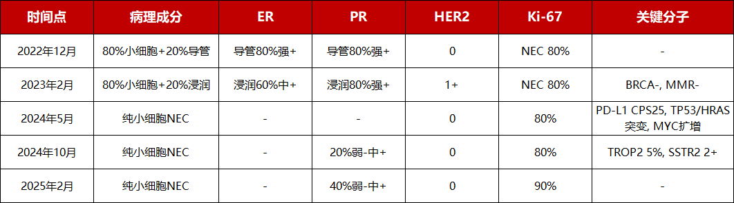
Postoperative pathology: invasive carcinoma of the left breast, with 80% small-cell carcinoma and 20% invasive carcinoma of no special type, grade 3. Immunohistochemistry: the small-cell carcinoma component was ER(−), PR(20% weak +), HER2(0), Ki-67(80%+), CD56(3+), Syn(3+); the ductal carcinoma component was ER(80% strong +), PR(80% strong +), HER2(0), Ki-67(60%+).
2. Radical Surgery (February 2023)
The patient underwent nipple-areola-sparing simple mastectomy of the left breast plus sentinel lymph node biopsy (0/4) at the Cancer Hospital of the Chinese Academy of Medical Sciences.
Postoperative pathology: 80% small-cell carcinoma + 20% invasive carcinoma of no special type, grade III (3+3+2=8 points), maximum tumour diameter 2.4 cm, involving skeletal muscle, with perineural invasion. Immunohistochemistry of the invasive carcinoma: ER(+, 60% moderate positive), PR(+, 80% strong positive), HER2(1+), Ki-67(80%); small-cell carcinoma: ER(−), PR(−), HER2(0), Ki-67(80%). Stage: pT2N0(sn). No BRCA1/2 or MLH1/2 mutations.
3. Postoperative Adjuvant Therapy (March 2023)
Adjuvant chemotherapy with etoposide + cisplatin (EP) combined with trilaciclib was planned for 4–6 cycles. Limited by severe myelosuppression, only 3 cycles were actually completed, and the planned standard course could not be finished. Disease-free survival (DFS) was only 2 months.
4. First Relapse and First-Line Therapy (May 2023 to December 2023)
The patient's tumour rapidly relapsed and fused into a mass. PET-CT on 23 August 2023 showed left anterior chest-wall metastasis and left axillary lymph node metastasis. First-line advanced therapy with goserelin + letrozole + abemaciclib (endocrine therapy combined with a CDK4/6 inhibitor) was given, with a progression-free survival (PFS) of only 3 months. From September 2023 left chest-wall radiotherapy was given, together with EP chemotherapy referring to pulmonary SCNEC. In December 2023 the left chest-wall tumour relapsed again with skin ulceration, and the patient switched to traditional Chinese medicine.
5. Local Intervention and Second-Line Therapy (April 2024 to October 2024)
On 29 April 2024 the patient underwent arterial infusion chemoembolization with albumin-bound paclitaxel 0.2 g + cisplatin 100 mg, and the chest-wall mass shrank.
On 21 May 2024 a chest-wall nodule biopsy was consistent with SCNEC. Genetic testing: PD-L1 CPS=25, TP53 and HRAS somatic mutations, MYC amplification. Imaging: multiple lymph node metastases in the left supraclavicular region, bilateral axillae, along the left internal mammary artery, the mediastinum and both hila; metastases in the left chest wall, lung and bone.
From 27 May 2024 second-line TP regimen (cisplatin 60 mg d1, d2 + albumin-bound paclitaxel 0.2 g d1, d8) combined with benmelstobart 1.2 g d8/q21d was given for 6 cycles. Partial response (PR) was achieved after 2 cycles (chest wall, lung and lymph nodes all regressed). Due to severe myelosuppression, cisplatin was stopped from cycle 5. Grade II peripheral neurotoxicity occurred. PFS was 4 months.
6. Third- to Fifth-Line Therapy (October 2024 to February 2025)
From 8 October 2024 third-line therapy with benmelstobart + anlotinib hydrochloride capsules was given for 1 cycle, with grade IV liver function impairment.
In November 2024 imaging showed comprehensive progression. Chest-wall pathology: SCNEC, ER(−), PR(20% weak-moderate +), HER2(0), Ki-67(80%), TROP2 (weak-moderate +, 5%), SSTR2 (2+).
On 11 November 2024 EC chemotherapy (liposomal doxorubicin hydrochloride injection) was given for 1 cycle and stopped because of adverse reactions.
Based on organoid drug sensitivity, on 9 December 2024 the regimen was changed to fifth-line therapy: benmelstobart + eribulin mesylate injection; because of febrile agranulocytosis, eribulin mesylate injection was adjusted to 2 mg d1,15/q28d. The last treatment was on 4 February 2025, with rapid progression of the chest-wall mass.
7. Sixth- and Seventh-Line Therapy (February 2025 to July 2025)
On 25 February 2025 sixth-line EC chemotherapy was given for 1 cycle (epirubicin hydrochloride injection 120 mg d1 + cyclophosphamide 900 mg d2/q21d); platelet count approached grade II reduction, and the chest-wall mass continued to enlarge.
On 11 March 2025 somatostatin receptor PET-CT showed multiple cystic-solid masses in the chest wall with high somatostatin receptor expression; multiple lymph node metastases in the right breast tissue spaces and axilla, left supraclavicular region and mediastinum; multiple bilateral lung metastases; T4 bone metastasis; left adrenal metastasis; and a small amount of left pleural effusion.
On 21 March 2025 seventh-line FOLFIRI was given: irinotecan hydrochloride injection 150 mg/m² (240 mg) d1 + fluorouracil 2.4 g/m² (3.9 g) civ 46 h/q14d.
On 10 April 2025 and 29 April 2025 chest-wall four-fraction radiotherapy was given (12 Gy/4 fractions/2 days and 14.8 Gy/4 fractions/2 days, respectively). During chemotherapy there was recurrent grade III-IV myelosuppression, febrile agranulocytosis and chest-wall soft-tissue infection, with wound secretion cultures growing carbapenem-resistant Pseudomonas aeruginosa and Enterobacter cloacae complex.
Because of extremely poor bone-marrow function, the fifth cycle (18 June 2025) was de-escalated to irinotecan hydrochloride injection monotherapy 240 mg d1 Q14d.
On 23 June 2025 follow-up CT showed enlargement of the left upper-lung pleura, right breast mass, lung and lymph nodes, with increased pleural effusion.
On 4 July 2025 PET-CT showed PD: the left chest-wall soft tissue still had tumour activity (range about 17 × 15 cm, SUVmax 2.6–5.8); new metastases in the right breast, right anterior chest-wall skin and soft tissue; multiple enlarged right axillary lymph nodes fused into a mass (largest 4.8 × 7.1 × 6.1 cm, SUVmax 5.4); a left upper-lobe perihilar nodule (diameter 2.4 cm, SUVmax 5.2) causing obstruction of the segmental bronchus; multiple left upper-lobe metastases; left diaphragmatic and paraspinal pleural metastases with a large left pleural effusion; and a new metastasis in liver segment IV (diameter 2.4 cm, SUVmax 6.4).
From 9 July 2025 to 30 July 2025 local radiotherapy to the right breast and right axilla was given, followed by oral traditional Chinese medicine.
8. Terminal State Before Admission
In the two weeks before admission the patient developed dyspnoea; chest CT showed a large left pleural effusion and she could not lie flat. Chest-wall cancer nodules increased and chest-back pain worsened, poorly controlled with fentanyl 8.4 mg q72h. She was in poor spirits, with poor appetite and low food intake, poor sleep, constipation, and a weight loss of 5 kg in the past month.
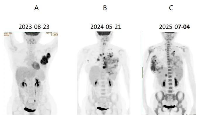
Final diagnosis: left breast cancer pT2N0(sn)M0 → M1, neuroendocrine carcinoma (triple-negative), with multiple metastases in the chest wall, lymph nodes, lung, liver, bone, pleura and adrenal gland.
II. Core Challenges
This case faced multiple challenges. NECB is relatively rare, and existing treatment evidence comes mainly from small-sample studies, lacking high-level evidence, so the clinical strategy must be formulated by combining the characteristics of both breast cancer and neuroendocrine carcinoma.
The patient had a ductal carcinoma component at initial diagnosis, but after relapse the tumour showed clear pathological evolution, and the original treatment strategy could not deliver sustained benefit. Post-relapse biopsy showed high Ki-67 expression, indicating increased tumour proliferative activity, suggesting possible phenotypic change during disease progression. Although molecular testing identified potential treatment leads such as PD-L1 expression, TP53/HRAS mutation and MYC amplification, after multiple lines of therapy the patient's bone-marrow reserve declined, with repeated severe myelosuppression and infection risk, further limiting subsequent antitumor treatment options.
At the same time, the patient developed a large chest-wall wound, recurrent infection, pleural effusion and pain in the advanced stage, and her overall state continued to decline. Under the dual influence of disease progression and declining treatment tolerance, the goal of care gradually shifted from purely controlling the tumour to balancing symptom relief and improvement of quality of life.
III. MDT Tiered Management and the End-Stage Decision
During disease progression the patient went through several key stages, including initial diagnosis and treatment, multi-line therapy after relapse, and end-stage treatment choices. Facing a rare pathological type, rapid disease progression and limited late-line options, the MDT team — bringing together the Breast Oncology Center, Pathology, Radiotherapy Center, Research Center, Diagnostic Radiology, Nuclear Medicine and Clinical Pharmacy — comprehensively assessed the disease characteristics, treatment responses and subsequent treatment possibilities at the patient's different stages.
Pathology changes after relapse: can the initial diagnosis still guide subsequent treatment?
Table 1: Pathological and molecular test results at different timepoints
★ Core finding:
The patient's pathology at different stages showed dynamic changes in tumour composition and receptor expression. At initial diagnosis there was a mixed pathological pattern, whereas after relapse the high-grade SCNEC component predominated, suggesting possible phenotypic change during disease progression.
★ Clinical caveat:
For patients with mixed NECB, pathology and molecular assessment should be repeated after relapse; subsequent treatment plans must not be based entirely on the initial specimen. Especially when the disease progresses rapidly or the treatment response does not meet expectations, repeat biopsy helps clarify the current tumour characteristics and provides a basis for subsequent treatment selection.
After identifying multiple potential targets, why is it still difficult to translate them into treatment benefit?
Multiple biopsies in this case showed dynamic changes in PD-L1, TROP2 and SSTR expression. The 2024 test showed PD-L1 CPS=25, suggesting a possible window for immunotherapy benefit, but subsequent treatment did not achieve sustained remission. Whole-body DOTATATE PET showed extensive SSTR positivity, providing a potential lead for PRRT, but evidence of efficacy in high-grade NEC is limited. The patient's TROP2 expression was low (5%), which may affect the benefit of TROP2 ADC drugs.
In addition, the DLL3-targeting drug Tarlatamab has shown some efficacy in heavily pretreated SCLC patients (DeLLPHI-301 study ORR 40%[1]), but its value in NECB is not yet clear. The CheckMate 032 study suggested that in SCLC, PD-L1 expression cannot effectively predict immunotherapy benefit[2], which also suggests that high PD-L1 expression does not necessarily mean the patient will benefit from immunotherapy.
Therefore, for such rare and highly heterogeneous tumours, the MDT needs to combine molecular test results, disease characteristics, treatment evidence and the patient's overall state to judge whether potential targets can truly be translated into treatment opportunities.
Can organoid drug-sensitivity results guide late-line treatment selection?
In the fifth-line stage, this case referred to the patient's tumour organoid drug-sensitivity test results in choosing eribulin mesylate injection. In-vitro testing suggested that eribulin mesylate injection and the EC regimen had some inhibitory effect on the tumour; after clinical application the patient's tumour progression was briefly controlled, but disease progression subsequently still occurred.
Tumour organoid drug sensitivity can provide a reference for late-line regimen selection, but in-vitro test results are not equal to the actual in-vivo treatment effect. Organoids can only observe the response of the drug acting directly on tumour cells, and cannot reproduce the patient's real state, such as immune status, concurrent infection, and impaired bone-marrow haematopoietic capacity after multiple rounds of chemotherapy — the complex realities in vivo. Therefore, organoid results alone cannot determine the treatment plan.
After multiple lines of therapy, how should treatment benefit be reassessed?
To clarify the patient's advanced treatment history, the MDT team sequenced the treatment lines according to the systemic antitumor regimens. Only full-body chemotherapy, endocrine therapy, targeted therapy and immunotherapy received in the advanced stage were counted as advanced systemic treatment lines; local radiotherapy, interventional procedures and symptomatic supportive care were not counted; and treatments in the postoperative adjuvant radical stage were not counted in the advanced treatment line sequence.
Table 2: The patient's complete advanced treatment lines
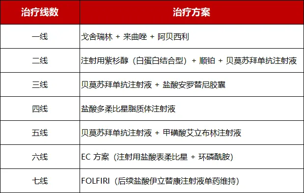
As the number of treatment lines increased, the patient experienced exploration of multiple treatment modalities, but the disease still continued to progress. After entering the late-line stage, the question the MDT focused on was no longer only "is there another drug to choose", but more importantly whether a new treatment plan could bring actual benefit, and whether the patient still had the foundation to continue treatment.
End-stage: continue looking for treatment opportunities, or adjust the goal of care?
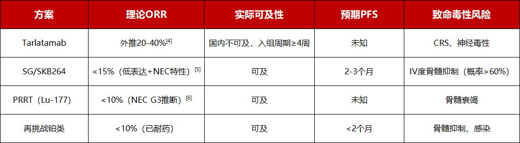
At the end-stage MDT in July 2025, all theoretical candidates were systematically reviewed: ADC (SG/SKB264, ASCENT study), multi-target TKI (cabozantinib, CABINET study)[3], DLL3 bispecific antibody (Tarlatamab, DeLLPHI-301), and HRAS inhibitor (tipifarnib, AIM-HN study).
However, no regimen could offer a clinically meaningful probability of benefit within a reasonable toxicity range. The patient's physiological reserve had fallen below the minimum safety threshold for any cytotoxic or targeted therapy: recurrent grade IV myelosuppression, uncontrolled multi-drug-resistant infection, cachexia, and ECOG 2-3. The 2011 international consensus by Fearon et al. defines refractory cachexia as increased catabolism, resistance to antitumor treatment, and expected survival of less than 3 months[7].
According to the principles of non-maleficence and beneficence in medical ethics, the certainty of harm from continuing antitumor treatment is far higher than the uncertainty of benefit[8]. ASCO guidelines recommend that palliative care be introduced within 8 weeks of diagnosis in patients with advanced cancer[9]. In this case, while the patient was conscious, a family meeting jointly decided to stop antitumor treatment and transition to palliative care.
IV. Clinical Insights
In this case, management faced multiple challenges including pathological heterogeneity, limited treatment options and multi-line drug resistance. At initial diagnosis the patient had mixed Luminal and neuroendocrine carcinoma components, and after disease progression the tumour gradually became predominantly high-grade neuroendocrine carcinoma, suggesting that the tumour may undergo phenotypic change under treatment pressure. Therefore, for patients with mixed NECB, when the disease relapses or the treatment response does not meet expectations, repeat biopsy and molecular testing should be performed promptly, rather than formulating subsequent plans entirely on the basis of the initial pathology. At the same time, multiple tests in this case identified potential treatment-related markers such as PD-L1, TROP2 and SSTR, but target expression did not fully translate into effective treatment opportunities, suggesting that precision treatment of advanced rare tumours not only requires finding targets, but also requires comprehensive judgment combining the disease type, evidence base and drug availability. In addition, organoid drug sensitivity can provide an auxiliary reference for late-line treatment selection, but it cannot fully simulate the patient's in-vivo immune status, infection factors and overall physiological changes after treatment, and test results still need to be interpreted in the clinical context.
As the disease entered the multi-line treatment stage, the focus of MDT decision-making also gradually shifted from "finding more treatment options" to "assessing treatment benefit". Although the patient still had theoretically available treatment options, after multiple lines of therapy she had developed severe myelosuppression, infection, cachexia and declining performance status, and further antitumor treatment was unlikely to achieve clear benefit. After comprehensive MDT assessment and full communication with the patient and family, the final decision was to stop antitumor treatment and transition to the palliative care stage. This case suggests that the management of advanced rare tumours requires attention not only to new drugs, new targets and new technologies, but also to dynamically adjusting the goal of care at different stages of the disease; when the risk of treatment gradually exceeds the potential benefit, supportive care centred on symptom control and improvement of quality of life is equally an important component of precision medicine.
Expert Commentary
Prof. Bai Li
Chair of the Academic Committee and Chair of the Ethics Committee, Beijing Arion Cancer Hospital; member of the International Committee on Challenging Tumours; formerly of the Department of Medical Oncology, First Medical Center of the Chinese PLA General Hospital; Chief Physician and Professor.
This is a high-quality case of whole-course management of a rare tumour, with notable strengths in the complete disease-evolution trajectory, tiered MDT and rational end-stage palliative-care decision. The main lessons centre on four aspects — early high-risk adjuvant therapy, regimen selection after relapse, rational use of organoids, and earlier timing of palliation — and carry good cautionary and reference value for clinical practice.
Although SCNEC arising in the breast is uncommon, apart from SCLC it can occur in every organ and tissue of the body and is encountered clinically. The first-line ORR of SCLC is high, reaching 70% or more, but once it fails, the second-line ORR drops precipitously; in particular, for platinum-resistant SCLC the second-line ORR with any chemotherapy drug is only 5%–15%, and even platinum-sensitive relapse is only 20%–30%. The mechanisms of resistance fall into five levels: intrinsic molecular plasticity of tumour cells, epigenetic resistance, tumour stem cells, the microenvironment, and host treatment-related damage.
Speaking of cellular phenotypic plasticity, this patient showed treatment-pressure-driven phenotypic drift: the 20% ductal carcinoma component of the primary lesion disappeared completely after relapse, replaced by a pure small-cell NEC clone. Epigenetically mediated chemotherapy resistance (predominantly non-genomic mutation) — such as epigenetic silencing of SLFN11, a key molecule for platinum/topoisomerase-inhibitor efficacy — and EZH2-mediated promoter methylation, histone modification, transcriptional reprogramming and upregulation of large numbers of resistance genes may be validated in basic theory or preclinical studies, but it is very difficult to detect the resistance-driving points directly by NGS. Tumour stem cells and drug efflux via MDR1/MRP have long been considered the main cause of chemotherapy resistance. In SCNEC the immune microenvironment is generally a "cold tumour"; even with PD-L1 positivity, overall tumour mutational burden is highly heterogeneous; there is massive infiltration of MDSC, Treg and tumour-associated macrophages, T cells struggle to infiltrate, PD-L1 cannot stably predict immune benefit, and the response rate of late-line single-agent ICI is extremely low.
Extrapulmonary SCNEC (EP-SCNEC, such as breast, cervical, gastrointestinal and genitourinary origin) has inferior efficacy to pulmonary primary SCLC. The reason is that although the pathological morphology is highly similar, the molecular origin, genomic characteristics and chemotherapy response differ significantly. Biologically, the genomic driver background differs: pulmonary SCLC has near-hallmark TP53 + RB1 double inactivation, whereas in EP-SCNEC the proportion of TP53/RB1 double inactivation decreases, with more HRAS, KRAS, PIK3CA and MYC amplification, and genomic differences among different primary organs are extremely large, with molecular heterogeneity far higher than pulmonary SCLC. The molecular basis of platinum response is not uniform, and some tumours are intrinsically insensitive to platinum. This breast NEBC case shows TP53 and HRAS somatic mutations and MYC amplification, which are typical extrapulmonary NEC features. The immune-microenvironment heterogeneity is stronger: the immune-combination benefit of pulmonary SCLC established by IMpower133 cannot be directly translated to EP-SCNEC; retrospective studies show that in EP-NEC, platinum + atezolizumab only improves ORR, without significant PFS or OS benefit; the immunogenicity of different organ origins differs greatly, and there is a lack of a unified immune-benefit marker.
From the clinical-reality perspective, extrapulmonary SCNEC treatment is in an evidence desert — there is no large phase III study, all are retrospective or case reports, and clinically one can only copy SCLC regimens, which is cross-tumour extrapolation without adequate validation.
The shortcoming in the discussion of this case is that early adjuvant therapy had a gap: the patient was pT2N0 but had skeletal-muscle involvement, perineural invasion and Ki-67 as high as 80%, belonging to the extremely high-risk group (perhaps the staging of SCNEC occurring in the breast should not follow the staging of breast ductal or invasive carcinoma), and only 3 cycles of EP were given before interruption due to myelosuppression — the inadequate adjuvant course is an important reason DFS was only 2 months. Regimen selection after first-line relapse had a trial-and-error element: after relapse the tumour was already predominantly pure SCNEC, yet Luminal-type endocrine therapy + CDK4/6 inhibitor was still chosen, with PFS of only 3 months and limited benefit. There was a misuse of organoid drug sensitivity: although the article points out the limitations of the in-vitro model, fifth-line therapy still relied heavily on organoid drug-sensitivity results in choosing eribulin mesylate injection, without fully weighting in-vivo infection, bone-marrow depletion and MYC/HRAS-driven proliferation, so there was in-vitro sensitivity but rapid in-vivo progression.
Despite the shortcomings in treatment, it is precisely the in-depth analysis and discussion of this rare type of breast cancer that gives us valuable experience, providing clinical information that can be referenced for similar patients encountered in future practice.
Prof. Sun Min
Chief Medical Officer, Beijing Arion Cancer Hospital.
From "what can we do" to "what should we do". During my residency training, my programme director once said: "Years ago, we asked ourselves, 'What can we do?' Today, we ask ourselves, 'What should we do?'" As medicine develops, the weight of this sentence becomes ever clearer. In the past, facing challenging tumours, we were often limited by the means of treatment; today, as new drugs, new targets and new technologies keep emerging, we need even more to judge: which choices are truly worth implementing for the patient in front of us?
This case of rare breast small-cell neuroendocrine carcinoma vividly presents the complexity of such decisions. A small-cell carcinoma component accounting for 80% at initial diagnosis suggests that treatment needs to focus on the aggressive component dominating the course. The subsequent repeat biopsies, molecular testing and organoid drug sensitivity reflect the team's effort to keep looking for opportunities when evidence is limited. However, the leads found by testing, the sensitivity observed in vitro, and the successful experience in other tumour types all need to be combined with the patient's pathological background and physical state to judge their actual value. When the disease keeps progressing and severe toxicity recurs, each line change requires re-weighing benefit, cost and the patient's wishes.
In observing the management of this case, another commendable detail is that the MDT team, while carrying out the main antitumor treatment, integrated nutritional support, acupuncture and traditional Chinese medicine intervention into whole-course care, striving to relieve suffering and maintain the patient's quality of life. This aligns with the concept of modern integrative oncology — patient-centred, within an evidence-based framework, combining appropriate adjunctive interventions with conventional oncology treatment to respond to the patient's physical and psychological needs throughout the disease course[10].
The key to such integration lies in selecting measures for specific symptoms according to the evidence, safety and patient preference of each measure, and continuously evaluating actual effects. For example, the joint SIO and ASCO guidelines have included acupuncture in the recommended management of some cancer-related pain[11]. For this case, nutrition, symptoms and quality of life were always given attention, giving "what should we do" a more positive and complete meaning: at every stage of striving to control the disease, helping the patient eat, suffer less and retain the ability to carry on daily life as much as possible.
As the condition developed, the MDT adjusting the focus of care to palliative care together with the patient and family became the continuation of this whole-course care philosophy. For a 42-year-old patient, such a decision is especially difficult, requiring full medical judgment and also a careful listening to her own expectations for the remaining time. The intensity of antitumor treatment can be adjusted, but care for the patient should always continue.
Medical progress gives us more and more "things we can do", and medical judgment helps us choose the "things we should do" for this patient. Among these are both treatment opportunities worth pursuing and every day worth carefully preserving.
Medical Disclaimer
This article is provided for general medical education and public health information only. It does not constitute medical advice, diagnosis, or treatment recommendations. Clinical outcomes depend on individual circumstances, and treatment decisions should always be made with a qualified physician.