
This article is a de-identified educational case review. It describes the clinical course of one patient managed at Beijing Arion Cancer Hospital and reflects the team's approach to assessing response during immunotherapy for biliary tract cancer. It is not medical advice, nor does it guarantee outcomes for other patients. Treatment decisions must be individualized by qualified physicians.
Case Overview: A 57-Year-Old Man with MSI-H Intrahepatic Cholangiocarcinoma
In June 2024, a 57-year-old man underwent radical surgery for left-sided intrahepatic cholangiocarcinoma: left hemihepatectomy with cholecystectomy, regional lymph node dissection, hepatic artery reconstruction, and hepaticojejunostomy.
Postoperative pathology reported intrahepatic cholangiocellular carcinoma, moderately to poorly differentiated, with vascular tumour thrombi and perineural invasion, and lymph node metastasis in 11 of 17 nodes dissected. Genomic profiling returned a tumour mutational burden (TMB) of 52.95 mutations per megabase together with microsatellite instability-high (MSI-H) status — the molecular profile of an immunologically "hot" tumour and one of the strongest predictors of benefit from immune checkpoint blockade.
One month after surgery, and before systemic treatment began, a left supraclavicular lymph node that had been present at baseline was biopsied. Pathology confirmed metastatic cholangiocarcinoma, and the patient was staged as pT2N1M1, stage IV.
Treatment therefore began in the setting of a resected primary with residual nodal and presumed micrometastatic disease, a very high TMB, and MSI-H status — a combination that made immunotherapy the logical backbone of systemic treatment.
The Core Challenge: A "Mixed Response" That Looked Like Treatment Failure
First line — lenvatinib plus pembrolizumab for six cycles. Restaging identified new intrahepatic nodules. Judged against conventional criteria these represented multiple new metastatic deposits, and the patient was assessed as having progressive disease (PD). The regimen was stopped.
Second line — regorafenib, gemcitabine, cisplatin, and cadonilimab for two cycles. Imaging then produced something far more confusing. Several intrahepatic lesions had disappeared entirely, yet the lesion in the lower segment of the right posterior hepatic lobe had enlarged markedly compared with the previous study. In other words, the liver looked simultaneously better and worse.
That mixed pattern framed the central dilemma of the case. In a patient receiving immunotherapy, does a new or enlarging lesion mean the tumour has escaped treatment, or does it mean the immune system is responding? If the team misread true progression, they would abandon a regimen that was working. If they misread true progression and continued anyway, they would lose valuable time in a disease with limited later-line options.

How the MDT Reasoned Through the Dilemma
The multidisciplinary team comprised medical oncology, hepatobiliary surgery, interventional therapy, radiology, and pathology. After two cycles of second-line treatment the assessment read "some lesions resolved, some lesions enlarged" — in other words, a mixed response arising from tumour heterogeneity. The discussion therefore narrowed to a single question: what is the nature of this enlarging lesion?
Perspective and Option A — Call It True Progression and Change Treatment Immediately
Rationale: Under traditional RECIST criteria for solid tumours, an enlarging lesion or a new nodule can be scored directly as disease progression, requiring the current regimen to be stopped at once and replaced with a salvage strategy.
Advantages: It terminates ineffective treatment promptly and avoids continued progression on a failing regimen.
Drawbacks: This patient belongs to an immunotherapy-favoured population (MSI-H, TMB-H) with a high probability of benefit, and most of his lesions had already responded. Judging response on imaging alone in this setting invites misclassification and would prematurely stop an effective immunotherapy regimen. In advanced intrahepatic cholangiocarcinoma, later-line options are limited, so an unnecessary switch would substantially narrow the patient's survival benefit.
Perspective and Option B — Call It Immune Pseudoprogression and Continue the Current Regimen
Rationale: Considering the patient's immunotherapy history, the overall reduction in tumour burden, and the atypical pattern of progression confined to a single isolated lesion, pseudoprogression was considered highly likely.
Advantages: If correct, the effective regimen can continue to control the tumour, maximising long-term immunotherapy benefit.
Risks: If the lesion really were true progression, continuing an ineffective regimen would permit rapid tumour growth and delay the best opportunity for effective treatment.
Perspective and Option C — Biopsy the Lesion to Guide an Individualised Decision
Rationale: Obtain a tissue diagnosis from the suspicious enlarging intrahepatic lesion so that the gold standard — pathology — determines its nature and directs subsequent treatment.
Advantages: It definitively separates true progression from immune-related inflammatory pseudoprogression, delivering a precise decision with no room for subjective misjudgement.
Risks: Biopsy carries a small risk of trauma, bleeding, and infection. In this case the lesion's location offered a safe needle path, so the overall risk was judged controllable.

The Decision: Biopsy the Enlarging Lesion
The team chose Option C. In the immunotherapy era, a new or enlarging lesion on imaging is not equivalent to true progression. This patient had an MSI-H/TMB-H "hot" tumour, and his atypical, isolated radiological progression after immunotherapy raised strong clinical suspicion of pseudoprogression rather than treatment failure.
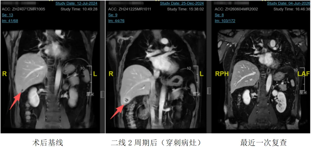
Biopsy of the enlarging intrahepatic lesion was performed promptly. Pathology reported abundant inflammatory cell infiltration with no tumour cells identified — an inflammatory nodule consistent with an immune-related reaction, confirming the diagnosis of immunotherapy pseudoprogression.
The pathological result became the anchor of the decision. Instead of switching regimens on an imaging finding, the team continued a treatment that was, in fact, working.
The same reasoning was later applied in retrospect to the first-line assessment: after six cycles of lenvatinib plus pembrolizumab, the "new intrahepatic nodules" may themselves have been pseudoprogression. Had the biopsy approach used during second-line treatment been applied then, the nature of those nodules might have been clarified earlier and the adverse effects of the subsequent quadruplet regimen avoided.
Treatment Pathway Executed
Radical left hemihepatectomy → first line lenvatinib + pembrolizumab × 6 cycles (assessed PD) → second line regorafenib + gemcitabine + cisplatin + cadonilimab × 2 cycles (mixed intrahepatic response: partial regression plus one enlarging lesion) → biopsy of the suspicious intrahepatic lesion → pathology: abundant inflammatory infiltration, no tumour cells — immune pseudoprogression confirmed → continue regorafenib + cadonilimab, stop the gemcitabine/cisplatin backbone → cadonilimab-related severe hypersensitivity and infusion reactions → switch to nivolumab + ipilimumab dual immunotherapy plus regorafenib → repeated imaging shows stable disease (SD) → current maintenance with nivolumab monotherapy plus reduced-dose regorafenib
Deep Dive on the Turning Point
The pivotal decision node in this case was the biopsy of the enlarging intrahepatic lesion. In the immunotherapy era, imaging findings of new or enlarging lesions do not automatically equate to true progression. This patient had an MSI-H/TMB-H "hot" tumour and developed isolated, atypical radiological "progression" on immunotherapy — a pattern in which pseudoprogression is clinically suspected. Timely biopsy confirmed an inflammatory nodule consistent with an immune-related reaction and established the diagnosis of immune pseudoprogression.
Managing the Unexpected: Toxicity and Regimen Adjustment
- Recognition and handling of pseudoprogression. When imaging showed an isolated enlarging lesion against an otherwise improving picture, the MDT did not blindly score PD; it pursued biopsy. The pathology result was the decisive evidence that spared the patient an unnecessary change of regimen and preserved the benefit of ongoing immunotherapy.
- Management of immune-related adverse events (irAEs). The patient later developed cadonilimab-related cutaneous hypersensitivity (rash with pruritus) and recurrent infusion reactions (hypotension, syncope). After safety reassessment, treatment was changed to a nivolumab plus ipilimumab dual-immunotherapy regimen. Under this regimen the rash remained controllable and infusions proceeded without incident.
Outcome and Follow-Up
Short-Term Efficacy
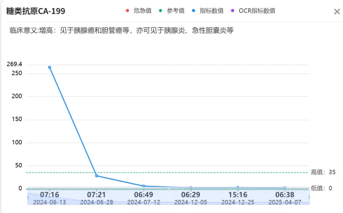
After pseudoprogression was confirmed and treatment was refined, the previously enlarging intrahepatic lesion showed no residual tumour activity, while earlier metastatic lesions continued to shrink and some resolved completely. Throughout the course no new distant metastasis has appeared, and repeated imaging assessments have consistently shown stable disease (SD) — durable and effective tumour control.
Survival and Functional Status
Since the second-line regimen was adjusted, the patient's disease has remained stable (SD) at every assessment. He has maintained an excellent functional state throughout treatment (ECOG performance status 0). During the quadruplet phase he experienced haematuria, hypertension, and rash, all of which resolved with supportive treatment; no grade 3 or higher severe adverse events occurred. After pseudoprogression was confirmed, his quality of life improved further, and he has been able to exercise and travel.
Ongoing Treatment and Follow-Up Plan
- Current regimen: nivolumab 480 mg every 4 weeks as maintenance therapy, with regorafenib reduced to 40 mg daily.
- Surveillance: upper abdominal MRI and tumour marker testing every 2–3 months, with particular attention to intrahepatic lesions and cervical lymph nodes.
- Plan if progression is suspected again: the MDT would again prioritise biopsy to distinguish true progression from pseudoprogression, and subsequent treatment would be based on the pathology and genomic results of that repeat biopsy.
Where This Case Sits in the Literature
Pseudoprogression is an increasingly recognised phenomenon in patients treated with immune checkpoint inhibitors (ICIs). It is defined as an initial increase in tumour size or the appearance of new lesions on imaging followed by shrinkage or stabilisation of the disease without any change in treatment — an unconventional response pattern that must be confirmed by repeat imaging [1]. Its pathological basis differs fundamentally from true progression: whereas true progression reflects proliferation of malignant cells, the enlarging lesion in pseudoprogression is characterised histologically by infiltration of T lymphocytes and other inflammatory cells, necrosis, or oedema, accompanied by an actual reduction in tumour burden [1].
Typical pseudoprogression usually presents as enlargement of existing lesions; the emergence of genuinely new lesions is considerably less common, particularly in tumours as immunotherapy-sensitive as MSI-H disease. Inflammatory pseudotumour is a special subtype of pseudoprogression and is clinically uncommon. In solid tumours such as non-small cell lung cancer (NSCLC), reported rates of pseudoprogression or inflammatory pseudotumour are on the order of 2–5%. These events most often occur early in the course of immunotherapy, although delayed presentations have also been reported [2].
At present the gold standard for diagnosing pseudoprogression remains pathological biopsy. In routine practice, however, clinicians often rely on a combination of iRECIST-based dynamic assessment, circulating tumour DNA (ctDNA) monitoring, and clinical dissociation — an improvement in symptoms that seems out of keeping with the radiological picture [3,4]. Where the distinction remains difficult, or where a vital organ's function is at stake, multidisciplinary discussion (MDT) is recommended.
For patients with MSI-H solid tumours who develop successive new lesions during immunotherapy, the possibility of an inflammatory pseudotumour should be considered. In suspected cases, and provided patient safety is assured, continuing treatment across the radiological finding while obtaining a prompt and safe biopsy is advisable. Determining the true nature of an enlarging lesion — tumour versus inflammation — is the key step in avoiding the premature termination of an effective treatment and achieving genuinely individualised care. This case provides a useful reference point for clinical decisions in comparable situations.
Expert Commentary
Prof. Bai Li
Chair, Academic Committee and Chair, Ethics Committee, Beijing Arion Cancer Hospital; Member, International Committee on Complex Oncology; formerly Department of Medical Oncology, First Medical Centre of the Chinese PLA General Hospital — Chief Physician, Professor
This is a comparatively rare individual case of MSI-H cholangiocarcinoma. The core clinical difficulty is this: during immunotherapy, imaging showed enlargement of an intrahepatic lesion with new nodules. Because the radiological appearance did not fit a typical metastatic deposit, yet metastasis could not be entirely excluded, the decision to question whether pseudoprogression was occurring — and to confirm it by biopsy — is highly commendable and worth recommending.
Immunotherapy should not be managed by blind adherence to conventional RECIST imaging criteria. Precise discrimination between true progression and post-immunotherapy pseudoprogression is essential, and biopsy pathology is the gold standard. Before proceeding to biopsy, however, one should first assess whether symptoms have improved, review laboratory indices, and evaluate imaging characteristics to judge whether the tumour is waxing or waning. Not every increase in tumour size requires biopsy — the incidence of pseudoprogression is, after all, low.
In routine practice, typical pseudoprogression consists mainly of enlargement of a single lesion; multiple new lesions are relatively uncommon. This patient instead presented with multiple suspicious new inflammatory nodules in the liver — a form of immune-related inflammatory pseudotumour that is rarely seen clinically. It has a very low incidence, most often appears early in immunotherapy, and is easily misdiagnosed as widespread progression. Comparable cases are uncommon, so the reference value of this case is relatively high: the MSI-H/TMB-H subtype of intrahepatic cholangiocarcinoma is itself uncommon, and multiple inflammatory pseudotumour-type pseudoprogression after immunotherapy is a rare presentation. It differs from conventional single-lesion pseudoprogression and adds clinical data on this particular scenario.
Prof. Sun Min
University of Pittsburgh Medical Center
This is a case report of considerable teaching value. In a rare subtype — MSI-H/TMB-H cholangiocarcinoma — the authors resisted the conventional RECIST reflex that "a new lesion means PD means switch lines" and instead used biopsy pathology as the gold standard to confirm an immune-related reaction. The decision chain is clear and reproducible, and the management of irAEs is sound and well judged. Particularly noteworthy is that the authors accurately distinguished classic pseudoprogression from the less common inflammatory pseudotumour/sarcoid-like reaction, and incorporated ctDNA into a comprehensive framework for assessing true versus pseudoprogression. These two points demonstrate a deep grasp of the complexity of response assessment in the immunotherapy era and make this article's mechanistic interpretation and clinical guidance clearly superior to that of an ordinary case report.
What This Case Illustrates
- In the immunotherapy era, be alert to pseudoprogression. For patients receiving immune checkpoint inhibitors — especially those in favoured populations such as MSI-H or TMB-H — the appearance of a new or enlarging lesion on imaging does not equal treatment failure. It must be interpreted together with the overall change in tumour burden and the lesion's radiological features, such as its margins and oedema.
- Biopsy is the gold standard for distinguishing true from false progression. When the imaging diagnosis is in doubt and the lesion can be safely accessed, pathological evidence should be actively obtained. In this case a single timely biopsy prevented the mistaken discontinuation of an effective immunotherapy regimen and directly altered the patient's prognosis.
- MDT is indispensable in complex decisions. Radiology contributed the atypical imaging features, medical oncology analysed the probability of immunotherapy benefit, and pathology guided the biopsy and diagnosis. Multidisciplinary collaboration produced the correct decision: confirm by biopsy, then continue treatment.
- Look back at earlier decision points. The first-line "new intrahepatic nodules" may also have been pseudoprogression. Applying the same biopsy-first logic earlier might have clarified the nature of those nodules sooner and avoided the adverse effects of the subsequent quadruplet regimen.
- Anticipate and manage irAEs. Prompt recognition of cutaneous hypersensitivity and infusion reactions to cadonilimab allowed a safe switch to a dual-immunotherapy backbone without losing disease control.
References
- Fountoukidis G, Tina E, Göthlin-Eremo A, Ullenhag G, Valachis A. Optimizing the clinical assessment of pseudoprogression in patients with solid tumors treated with checkpoint inhibitors: a systematic literature review and meta-analysis of associated features. Cancer Immunol Immunother. 2026 Jun 8;75(6):170.
- Fountoukidis G, Tina E, Göthlin-Eremo A, Ullenhag G, Valachis A. Optimizing the clinical assessment of pseudoprogression in patients with solid tumors treated with checkpoint inhibitors: a systematic literature review and meta-analysis of associated features. Cancer Immunol Immunother. 2026 Jun 8;75(6):170.
- Borcoman E, Kanjanapan Y, Champiat S, Kato S, Servois V, Kurzrock R, Goel S, Bedard P, Le Tourneau C. Novel patterns of response under immunotherapy. Ann Oncol. 2019 Mar 1;30(3):385-396.
- Persigehl T, Lennartz S, Schwartz LH. iRECIST: how to do it. Cancer Imaging. 2020 Jan 3;20(1):2.
Medical Disclaimer
This article is provided for general medical education and public health information only. It does not constitute medical advice, diagnosis, or treatment recommendations. Clinical outcomes depend on individual circumstances, and treatment decisions should always be made with a qualified physician.