This article is a de-identified educational case review. It describes the clinical course of one patient managed at Beijing Arion Cancer Hospital and reflects the team's approach to ultrasound-based differential diagnosis and biopsy targeting in a rare gynaecological tumour. It is not medical advice, nor does it guarantee outcomes for other patients. Treatment decisions must be individualized by qualified physicians.
Case Introduction
This issue analyses a rare case in which an elderly woman presented with dyspnoea and was found on imaging to have synchronous space-occupying lesions of the uterus and both adnexa, together with extensive metastases to the peritoneum, omentum and lymph nodes throughout the body. Her tumour marker CA125 was markedly elevated, and at first presentation the clinical picture strongly favoured an ovarian epithelial malignancy. Through multidisciplinary (MDT) assessment, discrimination of characteristic multimodal ultrasound features, and ultimately an ultrasound-guided biopsy, the team confirmed a rare primary uterine lymphoma. For difficult cases like this — synchronous uterine and adnexal involvement, unknown primary site, and a suspected gynaecological malignancy — the case provides a practical demonstration of ultrasound imaging-based differential diagnosis combined with precise MDT decision-making.
I. Case Overview: Baseline Status and Core Challenges
Baseline Status
A 78-year-old woman presented to an outside hospital on 20 May 2025 with two weeks of dyspnoea. Pelvic MRI showed multiple abnormal signal foci throughout the entire uterus and both adnexa, with lesions involving the bladder, rectum and vagina, accompanied by lymph node and peritoneal metastases. Abdominal CT showed peritoneal metastases, large-volume ascites and multiple enlarged retroperitoneal lymph nodes.
An indwelling peritoneal drain was removing approximately 1000 ml of ascites each day. The ascites cytopathology and PET-CT results were still pending, and she could not tolerate gastrointestinal endoscopy. Tumour markers were markedly elevated: CA125 538 U/mL and NSE 48 ng/mL.
She was transferred to Beijing Arion Cancer Hospital on 29 May 2025 for further diagnosis and treatment. She had no previous history of cancer; she was elderly and her general condition was frail.
Current Dilemmas
1. Diagnostic dilemma: synchronous multiple masses of the uterus and both adnexa, combined with extensive metastases to the peritoneum, omentum and multiple lymph nodes, made it impossible to distinguish the primary site or the nature of the lesions.
2. Therapeutic dilemma: with no definitive pathology and no evidence of the primary site, no precise systemic treatment plan could be formulated, and clinical management came to a standstill.
3. Decision dilemma: the patient was elderly, with massive ascites and a frail constitution, and could not tolerate laparoscopic invasive sampling. Obtaining tissue by minimally invasive puncture places extremely high demands on image localisation and target selection.
Key Assessment Results
Pelvic MRI showed multiple abnormal signal foci throughout the entire uterus and both adnexa, involving the bladder, rectum and vagina, with lymph node and peritoneal metastases. Abdominal CT showed peritoneal metastases, large-volume ascites and multiple enlarged retroperitoneal lymph nodes. The PET-CT result was pending. Clinically the picture favoured a malignancy of adnexal origin with widespread metastasis, and adnexal biopsy was recommended.
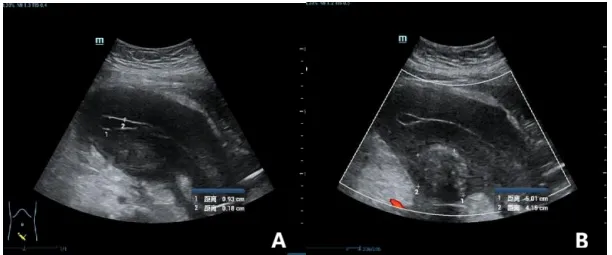
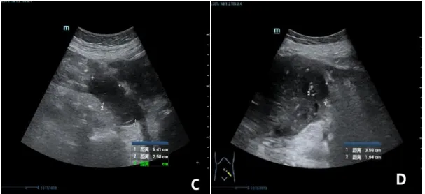
Ultrasound, however, showed diffuse uterine enlargement with overall hypoechoic myometrium, an intact endometrial line and no obvious focal mass effect; the bilateral adnexal hypoechoic masses had relatively regular borders. These findings differed markedly from the typical imaging features of ovarian epithelial cancer. Ultrasound considered primary uterine lymphoma most likely, with ovarian cancer to be excluded, and recommended revising the biopsy target.
II. Decision-Making: MDT Core Analysis and Strategy Deliberation
Treatment Pathway Options and Their Trade-offs
Option A: Perform an ovarian/adnexal lesion biopsy according to the initial judgement.
Rationale: The markedly elevated CA125, together with multiple masses of the uterus and adnexa and extensive peritoneal metastasis, fit the common clinical picture of ovarian epithelial cancer.
Limitations: If this is in fact a primary uterine lymphoma in which the ovaries are only secondarily involved, puncturing the adnexa is prone to a false-negative result, or may reveal only metastatic change.
Option B: Watch and wait, deciding only after the ascites cytopathology and PET-CT results are available.
Rationale: Rely on ascites cytology and the metabolic features of whole-body PET-CT to help establish the diagnosis.
Limitations: Ascites cytology has a limited positive yield and rarely permits precise subtyping; PET-CT can provide only a metabolic tendency and cannot replace pathology as the gold standard. Waiting would also delay the optimal treatment window in an elderly patient.
Option C: Ultrasound-guided targeted biopsy of the diffuse uterine lesion.
Rationale: Ultrasound showed a characteristic pattern — diffuse uterine enlargement, overall hypoechoic myometrium, no obvious mass effect, and a clear, intact, uninvolved endometrial line — consistent with the imaging features of lymphoma, making the uterus the suspected primary lesion.
Advantages: The biopsy is directed precisely at the suspected primary lesion, giving a high pathological yield and allowing the pathological subtype to be further defined. It is also minimally invasive and well tolerated, suiting a frail elderly patient.
Forming the Final Decision
The MDT team reached consensus: abandon the originally planned adnexal biopsy and instead perform an ultrasound-guided targeted biopsy of the diffuse hypoechoic area within the uterine myometrium. The aim was to obtain tissue from the suspected primary lesion with the least possible trauma, to establish the pathological type as quickly as possible, and to provide gold-standard evidence for onward specialist referral and systemic treatment.
Ultrasound-Guided Uterine Biopsy Pathology
(Diffuse hypoechoic area of the uterine myometrium) The biopsy tissue showed diffuse infiltration by medium-sized atypical lymphocytes. Combined with immunohistochemistry, this was considered non-Hodgkin diffuse large B-cell lymphoma, NOS, GCB type (not otherwise specified, germinal centre B-cell type).
Immunohistochemistry — A1: CD3 (T cells +), CD20 (+), CD30 (−), CD21 (−), Ki67 (MIB1) (+90%), CK (−), CD5 (T cells +), CD10 (+), Bcl-2 (+10%), Bcl-6 (+80%), c-MYC (+15%), CyclinD1 (−), PAX-5 (+), MUM1 (partially +), CD23 (−), CD19 (+), SOX-11 (−), TdT (−).
III. The Breakthrough: Treatment Course and Technical Points
Treatment Pathway Executed
Combined multimodal ultrasound assessment → MDT discussion revises the biopsy target → ultrasound-guided biopsy of the uterine lesion → pathology plus immunohistochemistry confirms lymphoma → bone marrow biopsy excludes marrow involvement → MDT formulates a targeted-immunotherapy plus chemotherapy regimen → 4.5 months of standard systemic treatment → PET-CT response evaluation and follow-up
Key Nodes Analysed in Depth
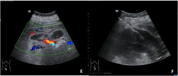
1. Technical points of combined multimodal ultrasound scanning. Transabdominal, transvaginal and transrectal scanning were combined to assess the overall morphology of the uterus, myometrial echogenicity, the endometrium, both adnexa, the pelvic peritoneum, the omentum, and the distribution, echogenicity and vascular features of superficial and deep lymph nodes throughout the body. The focus was on discriminating the characteristic ultrasound features of lymphoma — diffuse uniform hypoechogenicity of the uterus, overall enlargement without an obvious focal mass effect, and a continuous, intact endometrial line — from the typical imaging features of endometrial cancer, uterine sarcoma and ovarian epithelial cancer. This provided the core evidence for the clinical diagnosis and for selecting the biopsy target.
2. Precise targeting for ultrasound-guided puncture. Major vessels, bowel and areas of fluid were avoided. A solid, diffusely hypoechoic area within the myometrium was selected as the biopsy target, avoiding the benign fibroid nodule in the posterior wall. The procedure was fully visualised throughout, with real-time monitoring of the needle path — minimally invasive, safe and yielding reliable tissue, and well suited to an elderly patient with ascites and a poor general condition.
3. Specialist referral and formulation of systemic treatment. After ultrasound-guided uterine biopsy confirmed non-Hodgkin diffuse large B-cell lymphoma, GCB type, the patient was referred from gynaecology to the lymphoma medical oncology service. Based on stage IVb disease and a high-risk stratification, the MDT formulated a standard regimen of polatuzumab vedotin plus rituximab and mini-CHP, balancing efficacy against treatment tolerability in an elderly patient.
Short-Term Efficacy
After 4.5 months of standard systemic treatment, repeat PET-CT showed that the multiple lymph nodes in the retroperitoneum, mesentery and along both iliac vessels had shrunk markedly with no abnormal hypermetabolism; the metabolic activity of the thickened greater omentum and peritoneal lesions had returned entirely to normal; metabolism in the uterine lesion was markedly reduced; and the pelvic effusion had been completely absorbed. By the Deauville criteria the response was scored as CMR 1 — a complete metabolic response, representing a substantial treatment effect.
IV. What This Case Teaches
Diagnostic Thinking
When a patient presents with synchronous multiple masses of the uterus and adnexa together with an elevated CA125, it is easy to fall into an inertial diagnosis of ovarian cancer and to overlook rare entities such as primary uterine lymphoma. Ultrasound has a distinctive discriminating value here: diffuse uterine enlargement, overall hypoechogenicity of the myometrium, an intact uterine cavity structure and the absence of an obvious focal mass effect are important imaging clues to uterine lymphoma. Adnexal involvement usually appears as solid hypoechoic lesions with relatively well-defined borders — markedly different from the typically mixed cystic and solid appearance of ovarian epithelial cancer, with its papillary projections and infiltrative growth. The MDT model broadens the differential diagnostic thinking and prevents arbitrary diagnosis.
MDT Collaboration Model
This case fully demonstrates the core value of MDT in difficult and rare gynaecological tumours: it breaks down the limits of single-specialty thinking. The ultrasound department provided precise imaging characteristics and a suggested biopsy target; gynaecology, radiology, pathology and lymphoma medical oncology evaluated the case from multiple angles; and the diagnostic pathway and biopsy plan were corrected in time, avoiding blind treatment. In elderly, high-risk patients requiring minimally invasive management, MDT can close the loop across precision localisation, precision sampling, precision subtyping and precision treatment.
Expert Commentary
Prof. Wu Ming
Chief Physician, Professor and Doctoral Supervisor, Department of Obstetrics and Gynaecology, Peking Union Medical College Hospital
The patient's initial diagnosis was a high suspicion of ovarian cancer with widespread metastasis. The pre-set treatment approach was likewise to give neoadjuvant chemotherapy after the pathological diagnosis, followed by interval cytoreductive surgery, and then to continue chemotherapy after surgery. In this case, however, the diagnosis changed as a result of the ultrasound-guided needle biopsy — the eventual pathology was lymphoma. That made an enormous difference to the patient's actual benefit.
The plan had originally been to proceed with surgery as for advanced ovarian cancer, but the result of the ultrasound-guided needle biopsy changed the entire direction of treatment. For advanced cancer this amounted to a fundamental change in management. It avoided the trauma of surgery while also meeting the contemporary pursuit of precision oncology.
Prof. Wang Jingwen
Chief Physician, Lymphoma Centre, Beijing Arion Cancer Hospital
This case fully illustrates the role of ultrasound-guided needle biopsy in the diagnosis of lymphoma. First, the gold standard for confirming lymphoma is histopathology together with immunohistochemistry. Second, ultrasound-guided puncture is the most commonly used and most minimally invasive means of obtaining tissue from a lesion, and is particularly suitable as an alternative to surgical excision when the lesion cannot be resected — for example deep or multiple lesions, or widespread generalised lymphadenopathy where no surgical biopsy is feasible — as well as in elderly, frail patients with poor coagulation or underlying disease who cannot tolerate general anaesthesia or surgical excisional biopsy. In addition, ultrasound-guided puncture requires only local anaesthesia, with minimal trauma, rapid recovery and a lower risk of sampling.
This patient was elderly and frail, with multiple comorbidities and widespread generalised lymphadenopathy. It was precisely because of a timely ultrasound-guided biopsy that the pathology was confirmed as non-Hodgkin diffuse large B-cell lymphoma, GCB type, and that, after MDT, a regimen suited to her was devised according to tumour stage and prognostic stratification, balancing efficacy against tolerability in the elderly — achieving a very good response. Our ultrasound department has accumulated extensive experience in puncture and has played a very important role in accelerating the diagnosis and differential diagnosis of tumours, greatly benefiting other clinical departments and patients alike.
Medical Disclaimer
This article is provided for general medical education and public health information only. It does not constitute medical advice, diagnosis, or treatment recommendations. Clinical outcomes depend on individual circumstances, and treatment decisions should always be made with a qualified physician.