This article is a de-identified educational case review. It describes the clinical course of one patient managed at Beijing Arion Cancer Hospital and reflects the team's approach to complex chest wall sarcoma. It is not medical advice, nor does it guarantee outcomes for other patients. Treatment decisions must be individualized by qualified physicians.
Case Overview: A Tumor That Doubled in Two Months
In late 2025, a 69-year-old man sought care for a mass on the left chest wall that had been present for two months and was steadily enlarging. Enhanced chest CT performed at another hospital raised suspicion of a malignant left chest wall lesion. Chest MRI described a soft-tissue mass measuring approximately 7.5 × 5.5 cm, involving the latissimus dorsi and serratus anterior, with the adjacent ribs still appearing continuous.
Whole-body staging with PET-CT changed the picture. It showed confluent nodules and masses in the left chest wall, the largest approximately 8.6 × 5.4 cm, with markedly elevated FDG uptake and a SUVmax of 46.3 — a level consistent with a highly aggressive malignancy. Importantly, no distant metastasis was identified.
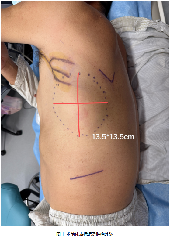
At Beijing Arion Cancer Hospital, ultrasound performed in October 2025 measured a solid mass of 9.2 × 8.3 × 4.8 cm within the chest wall musculature of the left hypochondrium. It was ill-defined and irregular with high-resistance internal blood flow — an appearance suggesting sarcoma. In other words, within roughly one month of imaging the lesion had grown again.
Percutaneous biopsy was performed in early October 2025 and reported a malignant mesenchymal tumor. Pathology review at Arion on 13 October 2025 established the diagnosis of dedifferentiated leiomyosarcoma. Immunohistochemistry showed diffusely positive vimentin, focally positive smooth muscle actin (SMA), and a Ki-67 proliferation index of approximately 70%, with negative desmin, S-100, and CD34. Taken together, these findings described a tumor that was high grade, proliferating rapidly, locally aggressive, and at high risk of recurrence.
Why This Case Was Difficult
- Rapid growth and wide invasion. The tumor had grown from about 5 cm to nearly 10 cm in two months, with infiltrative, expansile growth involving the latissimus dorsi and serratus anterior and crossing three ribs. Its relationship to the 7th and 8th ribs was close, with suspected periosteal involvement.
- Major structural loss requiring reconstruction. Resecting three ribs would remove a substantial part of the chest wall's supporting frame, risking flail chest, paradoxical respiration, and respiratory failure. Because the latissimus dorsi was itself involved, it could not serve as a flap; primary closure under excessive tension would risk necrosis, infection, and wound dehiscence.
- Advanced age with significant comorbidity. At 69 years old, the patient also had hypertension, coronary and aortic atherosclerosis, fatty liver disease, and a history of thyroid surgery — all of which raised perioperative cardiopulmonary risk.
- A histology that resists chemotherapy and radiotherapy. Dedifferentiated leiomyosarcoma is a high-grade subtype with poor responses to conventional systemic treatment. Complete surgical removal with reconstruction remained the only potentially curative path.
MDT: Where the Decisions Were Made
An multidisciplinary team (MDT) was convened, bringing together thoracic surgery, plastic and reconstructive surgery, medical oncology, radiology, ultrasound, pathology, nuclear medicine, radiation oncology, anesthesiology, the operating room team, and critical care. External specialists in thoracic medical oncology from Beijing Chest Hospital and in thoracic surgery and plastic surgery from Peking Union Medical College Hospital contributed to the discussion.
The team reviewed the history — rapid growth, broad invasion, involvement of the latissimus dorsi and part of the serratus anterior, and a tumor crossing three ribs — and settled on a single unified diagnosis: giant dedifferentiated leiomyosarcoma of the left chest wall.
Two decisions followed.
First, no neoadjuvant treatment. Dedifferentiated leiomyosarcoma responds poorly to chemotherapy and radiotherapy. Because the tumor was progressing rapidly, giving preoperative treatment would risk losing the surgical window. The team recommended proceeding directly to surgery in pursuit of R0 resection, with adjuvant treatment planned afterward according to pathology and genomic findings.
Second, extended resection with chest wall reconstruction was mandatory. The plan required adequate margins including the overlying skin; intraoperative assessment of periosteal involvement with resection of two to three ribs as needed; rigid reconstruction of the bony chest wall using a titanium plate with polypropylene mesh reinforcement; and soft-tissue coverage. Because the latissimus dorsi was involved and unusable, the plastic surgery team proposed a rectus abdominis musculocutaneous flap or an adjacent flap to close the large defect. The team also explained the risk of injury to the long thoracic and thoracodorsal nerves, which could cause winged scapula, restricted shoulder movement, and sensory changes.
Options Weighed: Three Strategies
Option A — simple excision with primary closure. Inadequate margins would carry a high risk of recurrence; loss of chest wall support would risk paradoxical respiration and pulmonary complications; and wound breakdown under tension was highly likely.
Option B — palliative resection with radiotherapy. This could not achieve cure, would likely leave residual tumor, and offered limited survival benefit in a rapidly progressing disease.
Option C — MDT-planned wide resection with chest wall reconstruction. Adopted. It offered complete tumor removal with a safe margin (R0), restoration of a rigid chest wall to eliminate respiratory compromise, one-stage reconstruction of a massive soft-tissue defect by the plastic surgery team, and the best achievable balance between recurrence control, function, and quality of life.
Reconstruction Planning: Ribs, Titanium Plate, Mesh, and Free Flap
Each discipline contributed a specific piece of the plan. Thoracic surgery defined the extent of resection and the number of ribs to be removed, recognizing that multi-rib resection would render the chest wall unstable and that titanium plate reconstruction with mesh reinforcement was essential. Plastic and reconstructive surgery designed a free flap to guarantee blood supply and reliable coverage, since the latissimus dorsi could not be used. Pathology, radiology, and nuclear medicine confirmed the grade, margins, and absence of metastasis that justified curative-intent surgery. Anesthesiology, critical care, and the operating room team planned for a long, complex, multidisciplinary procedure. Medical oncology and radiation oncology mapped out postoperative adjuvant options to reduce late recurrence.
The final unified plan was: wide radical resection of the chest wall tumor + resection of three ribs + titanium plate chest wall reconstruction + polypropylene mesh reinforcement + free flap soft-tissue reconstruction with the plastic surgery team.
Surgery and Recovery
The tumor was removed en bloc with an extended safe margin, clearing the involved musculature completely. The three involved ribs were resected as planned. A titanium plate restored the rigidity of the thoracic cage, and polypropylene mesh reinforced the repair, eliminating paradoxical movement. Because the latissimus dorsi was involved, the plastic surgery team joined the operation to perform a free flap transfer, restoring blood supply, covering the defect, and improving the contour of the chest wall. Closure was then performed in careful layers with drains left in place to support primary healing.
The postoperative course was uneventful. Vital signs and respiratory function remained stable with no paradoxical respiration, pneumonia, flap necrosis, pleural effusion, or pneumothorax. The incision healed primarily with a satisfactory contour and a stable, intact chest wall, and the patient moved on to adjuvant treatment planning.
Pathology confirmed that both the skin and deep margins were free of tumor — an R0 resection. Follow-up is scheduled with chest imaging at one month, genomic profiling to inform individualized adjuvant therapy, and ongoing surveillance of tumor recurrence, titanium plate position, and flap healing.
Where This Tumor Sits in the Literature
Primary dedifferentiated leiomyosarcoma of the chest wall is an extremely rare high-grade soft-tissue sarcoma, accounting for fewer than 4% of all chest wall soft-tissue sarcomas. It most often arises from the intercostal muscles, latissimus dorsi, or fascial mesenchymal tissue, and it is more common in middle-aged and older men, with a median age at diagnosis of about 64 years — closely matching this 69-year-old patient. Large retrospective series indicate that chest wall dedifferentiated leiomyosarcoma behaves considerably worse than its counterparts in the limbs or retroperitoneum: tumor doubling time is frequently shorter than three months, the Ki-67 index is commonly above 50%, and the risks of local invasion and early distant spread are substantially higher (Journal of Surgical Oncology, 2024).
Because the disease is so rare, no single standardized guideline exists. Treatment is usually individualized by combining soft-tissue sarcoma recommendations from NCCN and CSCO with the specific anatomy of the chest wall, and an MDT approach has become the preferred model for complex chest wall sarcoma. Imaging-based risk stratification comes first: a high SUVmax (above 10), a maximum diameter greater than 8 cm, and multi-muscle invasion all point to an aggressive phenotype. This patient's SUVmax of 46.3 with a mass approaching 10 cm placed him firmly in the high-risk category.
Some centers recommend preoperative neoadjuvant chemotherapy (doxorubicin plus ifosfamide) or concurrent radiotherapy for large high-grade soft-tissue sarcomas in the hope of shrinking the tumor and improving R0 rates. However, two multicenter retrospective cohort studies (Annals of Thoracic Surgery, 2025) reported objective response rates to cytotoxic chemotherapy of only 11–16% in dedifferentiated leiomyosarcoma, with limited local control benefit from radiotherapy — and in rapidly progressing patients, preoperative systemic therapy can delay the surgical window and may increase the risk of distant metastasis. That evidence is consistent with this team's decision to forgo neoadjuvant treatment. Only in patients whose tumors encase major vessels and cannot be resected in one stage might targeted therapy combined with chemotherapy be cautiously attempted.
For adjuvant treatment, the high risk of recurrence argues for postoperative radiotherapy. Leiomyosarcoma is also among the relatively chemotherapy-sensitive subtypes of soft-tissue sarcoma. Sarculator risk assessment predicted a 10-year overall survival of 30–40% for this patient — well below the 60% threshold — placing him in the group that benefits from chemotherapy (disease-free survival hazard ratio 0.49; overall survival hazard ratio 0.50). Depending on age and comorbidity, adjuvant chemotherapy can reasonably be discussed. If chosen, the AD regimen (doxorubicin 75 mg/m² plus dacarbazine 750–1000 mg/m²) is preferred for its favorable activity and tolerability in leiomyosarcoma compared with AIM, given for three to four cycles and sequenced with radiotherapy, with close cardiac monitoring.
What This Case Illustrates
- Pathology first, then strategy. Preoperative biopsy and multidisciplinary pathology review agreed closely with the final surgical specimen, confirming a dedifferentiated leiomyosarcoma with a high Ki-67 index. That diagnosis is what ruled out preoperative systemic treatment and protected the surgical window.
- Imaging defines the operation. Ultrasound and chest MRI delineated the tumor's borders and its relationship to the latissimus dorsi and serratus anterior, and correctly flagged the involvement of multiple ribs. Imaging predictions matched intraoperative findings and final pathology, directly supporting the decision to perform a large en bloc resection with single-stage reconstruction.
- Reconstruction is planned before the resection begins. Titanium plate, polypropylene mesh, and microvascular anastomosis instruments were prepared in advance, and operating room scheduling was organized around a long, complex procedure.
- Technique protects the outcome. Meticulous flap design, tension control, and preservation of blood supply were decisive for primary healing; early ligation of intercostal arterial branches reduced bleeding and kept the operative field clear.
- MDT closes the loop. The team unified the diagnosis, rejected ineffective therapy, designed the resection and reconstruction in detail, anticipated nerve injury and respiratory complications, and coordinated management from preoperative assessment through surgery, recovery, and adjuvant treatment.
Expert Commentary
Prof. Li Li, Peking Union Medical College Hospital
This 69-year-old patient with a giant dedifferentiated leiomyosarcoma of the chest wall presented the classic features of this disease: rapid growth, aggressive local invasion, high-grade histology, and poor sensitivity to chemotherapy and radiotherapy. The entire course was guided by multidisciplinary discussion with external experts, and the decisions were precise — making this a textbook example of curative resection with single-stage chest wall reconstruction in a high-risk, complex chest wall tumor.
Two points deserve emphasis. First, the preoperative biopsy and multidisciplinary pathology review aligned closely with the final surgical pathology, confirming dedifferentiated leiomyosarcoma with a high Ki-67 index and clarifying the tumor's aggressive biology. Because this subtype responds poorly to conventional chemotherapy and radiotherapy and progresses very quickly, the team's decision to place R0 surgical resection at the center of treatment — rather than pursuing preoperative neoadjuvant therapy that would have delayed the optimal surgical window — rested on solid pathological grounds.
Second, preoperative ultrasound and chest MRI proved their value. They defined the tumor's borders and its infiltrative relationship with the latissimus dorsi, serratus anterior, and other intercostal muscles, and correctly indicated involvement of multiple ribs with widespread destruction of normal chest wall structure. These predictions matched intraoperative exploration and final pathology, directly justifying a large en bloc chest wall resection with single-stage reconstruction.
Perioperative logistics also mattered. Titanium plates, polypropylene mesh, reconstructive materials, and microvascular anastomosis instruments were prepared in advance according to the MDT plan, with equipment and operating room scheduling arranged for a high-complexity reconstruction — an essential precondition for a long, multidisciplinary operation to proceed smoothly.
The operation followed the MDT plan exactly: wide R0 resection of the chest wall tumor, en bloc removal of three involved ribs, rigid reconstruction with titanium plate and mesh reinforcement, and simultaneous free flap soft-tissue reconstruction with the plastic surgery team. In repairing such a large chest wall defect, the design of the flap donor site and surrounding incisions, tension control, and preservation of blood supply determine success; here the incision layout and flap contouring created the conditions for primary healing and reliable flap survival. Early ligation of intercostal arterial branches reduced intraoperative and field bleeding and kept the dissection planes clear.
Recovery was smooth and orderly. Vital signs and respiratory function stayed stable, with no paradoxical respiration, chest wall collapse, pulmonary infection, or flap necrosis; drain volumes and character were as expected and drains were removed at the appropriate time. Both the incision and the free flap healed primarily, with satisfactory chest wall contour, stability, and functional recovery — allowing the patient to transition to individualized adjuvant treatment. Final pathology showed no tumor at the skin or deep margins, confirming R0 resection. Taken together, this case demonstrates how precise pathological classification, accurate imaging assessment, scientific multidisciplinary decision-making, thorough preoperative preparation, meticulous surgical technique, and multidisciplinary reconstruction can achieve tumor eradication, restoration of chest wall structure, soft-tissue closure, and preservation of function and appearance in an elderly patient with a giant, highly malignant chest wall sarcoma.
Medical Disclaimer
This article is provided for general medical education and public health information only. It does not constitute medical advice, diagnosis, or treatment recommendations. Clinical outcomes depend on individual circumstances, and treatment decisions should always be made with a qualified physician.