BEIJING — In this extraordinary case — the fifth volume of Arion's "Case Breakthrough Notes" (破局手记) series — a 23-year-old man presented with an extremely rare, newly defined entity in the 2021 WHO Classification of Tumors of the Central Nervous System: Primary Intracranial Sarcoma, DICER1-Mutant (WHO Grade 4). Approximately 87% of cases occur in children and adolescents, making adult cases extraordinarily rare, with virtually no high-level evidence to guide treatment. What followed was a testament to the power of multidisciplinary collaboration, molecular pathology, and evidence-based improvisation in the face of an unknown clinical frontier.
The Case: A Young Man with a Brain Tumor Nobody Recognized
The patient, a 23-year-old male with no significant medical history or family history of cancer, presented with bilateral lower extremity sensory abnormalities and intermittent claudication for over one month, accompanied by intermittent headaches for two weeks. An outside hospital's contrast-enhanced brain MRI revealed multiple right parietal lobe masses (largest ~3.7 cm), adjacent to the dura, with prominent homogeneous enhancement and a dural tail sign, high signal on DWI, and compression of the right lateral ventricle with surrounding edema. Initial differential diagnosis: primary central nervous system lymphoma.
A stereotactic biopsy was performed at the outside hospital. Pathology was inconclusive: "spindle cell malignant tumor components, favoring malignant mesenchymal origin; primary intracranial sarcoma with DICER1 mutation cannot be excluded." The case was referred to Beijing Arion Cancer Hospital for definitive diagnosis and treatment.
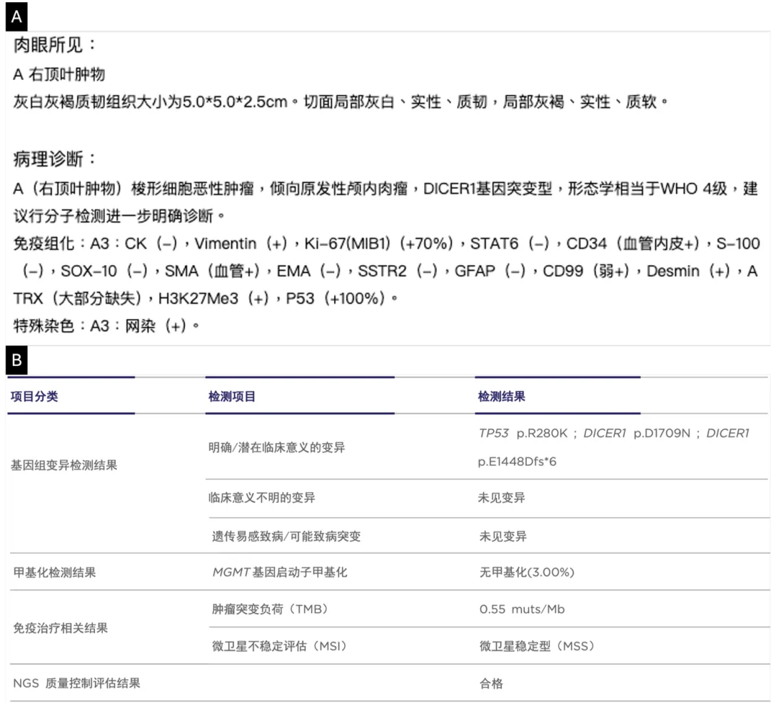
Stereotactic biopsy pathology report: spindle cell malignant tumor, favoring mesenchymal origin; primary intracranial sarcoma with DICER1 mutation could not be excluded. The diagnostic challenge was immediately apparent.
Three Core Challenges: Rare, No Guidelines, High Risk
The MDT team — comprising Neuro-oncology Center, Tumor Rehabilitation Center, Lymphoma Center, Radiation Oncology, Pathology, and Diagnostic Imaging (Radiology and Nuclear Medicine) — identified three critical challenges:
Challenge 1: Pathological diagnosis is extraordinarily difficult
Primary intracranial sarcoma, DICER1-mutant, was added as a new entity in the 5th edition (2021) of the WHO Classification of CNS Tumors. Approximately 87% of cases occur in children and adolescents. Adult cases are exceedingly rare, with extremely limited published data. The tumor's diverse mesenchymal differentiation features make it nearly impossible to distinguish from glioma or lymphoma on limited biopsy specimens. Definitive diagnosis requires dual verification: morphology + molecular pathology.
This patient's Ki-67 proliferation index was 70–80%, indicating extremely aggressive tumor biology with high risk of rapid progression and recurrence.
Challenge 2: No evidence-based treatment guidelines exist
Global evidence for this entity comes entirely from small case reports or retrospective series. No prospective clinical trials in adults have been conducted. Treatment strategy must be extrapolated from soft tissue sarcoma experience combined with CNS tumor biology — requiring the MDT team to essentially create a treatment pathway from scratch, guided by the closest available evidence.
Challenge 3: The surgical dilemma — GTR vs. functional preservation
The tumor was located in the right parietal lobe, adjacent to sensory and motor cortices. Gross total resection (GTR) is the strongest prognostic factor, but achieving it risks catastrophic neurological deficits. The team had to navigate the narrowest of margins: maximal tumor removal while preserving the patient's ability to walk, feel, and live independently.
MDT Decision: Creating Evidence from Nearest Neighbors
Surgery — Gross Total Resection (GTR): The Cornerstone
A 2022 systematic review of 52 cases showed that GTR was the strongest independent prognostic factor for primary intracranial sarcoma (multivariate Cox regression: HR = 0.061, p = 0.002). Patients who received GTR plus radiation therapy had a 2-year overall survival rate of 66.3%. Adult patients treated with surgery combined with ICE chemotherapy and radiation achieved a median OS of approximately 30.8 months — significantly better than subtotal resection or biopsy alone.
Decision: GTR is the non-negotiable foundation. The surgery would be performed by a neurosurgeon with extensive experience in neuro-oncology, using neuronavigation and intraoperative electrophysiological monitoring to protect motor cortex and venous sinus structures.
Adjuvant Therapy — ICE Chemotherapy + Concurrent Radiation
The ICE regimen (ifosfamide + carboplatin + etoposide) has demonstrated the most consistent efficacy in pediatric intracranial sarcoma cases. Given the tumor's basal attachment to the meninges (with partial blood-brain barrier disruption) and postoperative MRI showing neovascularization signals, the MDT team judged that systemic chemotherapy would be accessible. Radiation target dose: PTV 60 Gy in 30 fractions, starting 4 weeks post-surgery.
Decision: Postoperative ICE chemotherapy + concurrent radiation, starting within 4 weeks of surgery.
Exclusion of Other Options
Temozolomide (TMZ): The patient's MGMT promoter was unmethylated (3% methylation rate). TMZ benefit probability is extremely low in this molecular context. Excluded.
MEK inhibitors (MEKi): Approximately 77% of primary intracranial sarcomas carry RAS/RAF/MAPK pathway mutations, making MEKi a theoretical option. However, this patient's NGS revealed wild-type status for KRAS, NRAS, and BRAF. Excluded.
The Treatment Journey: From Diagnosis to Control
Preoperative Workup and Surgery
Preoperative evaluation revealed elevated liver enzymes (ALT 160.3 U/L), requiring hepatic protection before surgery. Under general anesthesia, the patient underwent right parietal craniotomy with supratentorial multiple mass excision, achieving GTR.
Postoperative pathology and molecular diagnosis confirmed the definitive diagnosis:
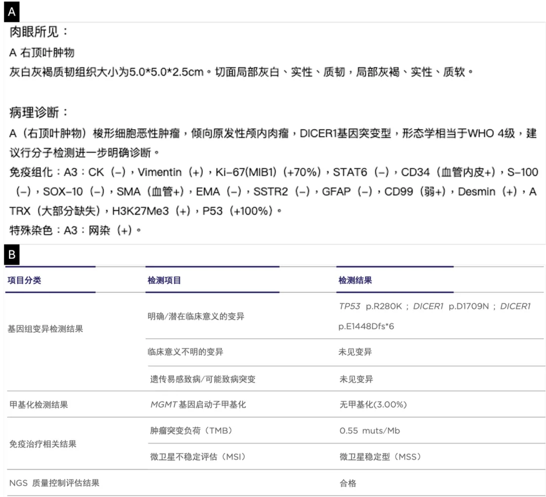
Postoperative pathology and NGS report confirming primary intracranial sarcoma, DICER1/TP53 co-mutation. (A) Pathology report with immunohistochemistry results; (B) NGS genetic testing report showing DICER1 and TP53 mutations.
Final diagnosis: Primary intracranial sarcoma, DICER1/TP53 co-mutant.
Immunohistochemistry:
- Vimentin (+)
- Desmin (+)
- Ki-67 (+70%)
- P53 (+100%)
- GFAP (-)
- S-100 (-)
- ATRX: mostly absent
NGS molecular testing:
- DICER1: p.D1709N and p.E1448Dfs*6 (biallelic inactivation)
- TP53: p.R280K
- MGMT promoter: unmethylated (3%)
- RAS/RAF/MAPK pathway: wild-type (KRAS, NRAS, BRAF)
Intraoperative Findings and Technical Challenges
Intraoperative exploration revealed multiple tumors located in the right parietal parasagittal region and deep parietal lobe. The tumor tissue was soft and firm in heterogeneous consistency, with extremely rich vascularity, intratumoral hemorrhage, and poorly defined boundaries with surrounding brain tissue. Through meticulous microsurgical dissection under neuronavigation guidance and electrophysiological monitoring, the surgical team strictly protected the motor cortex and venous sinus, gradually dissected the tumor, and overcame intraoperative bleeding, unclear margins, and the risk of functional zone injury — ultimately achieving microscopic gross total resection (GTR).
Postoperative Treatment: Radiation and ICE Chemotherapy
Approximately 4 weeks post-surgery, the patient started radiation therapy: intensity-modulated radiation therapy (IMRT) to the surgical bed, PTV 60 Gy in 30 fractions. Concurrently, ICE chemotherapy was initiated:
- Ifosfamide (IFO): 1.2–1.5 g/m², Days 1–3
- Carboplatin (CBP): AUC = 5, Day 1
- Etoposide (VP-16): 100 mg/m², Days 1–3
- Cycle interval: Every 3 weeks
As of the latest follow-up, the patient has completed 5 cycles of ICE chemotherapy according to the planned 8-cycle protocol.
Peri-treatment Complication Management
Hepatic injury: Baseline moderate fatty liver disease with elevated ALT (up to 210 U/L). Managed with hepatic protection therapy; controlled to acceptable levels before chemoradiation initiation.
Bone marrow suppression: Grade III myelosuppression occurred before Cycles 2 and 4 (WBC nadir 2.24 × 10⁹/L, neutrophil nadir 0.47 × 10⁹/L). Managed with G-CSF support (WBC recovered to 7.20 × 10⁹/L, neutrophils to 5.66 × 10⁹/L). Prophylactic long-acting G-CSF was initiated from Cycle 2 onward.
Urinary tract infection: Morganella morganii subsp. morganii infection identified before Cycle 4 (urine WBC 3+). Targeted anti-infective therapy achieved clearance; subsequent treatment continued uninterrupted.
Outcome: Functional Recovery and Tumor Control
Functional status: At the time of the 5th cycle ICE completion, the patient's preoperative lower extremity sensory abnormalities and claudication had significantly improved compared to initial admission. Activities of daily living are fully preserved.
Imaging follow-up: Postoperative 3-month contrast-enhanced brain MRI showed the surgical cavity with linear and lace-like enhancement, significantly reduced compared to the 1-week postoperative early imaging. Enhancement range and perilesional edema were markedly decreased, with no clear evidence of tumor recurrence — indicating effective treatment and good local control.
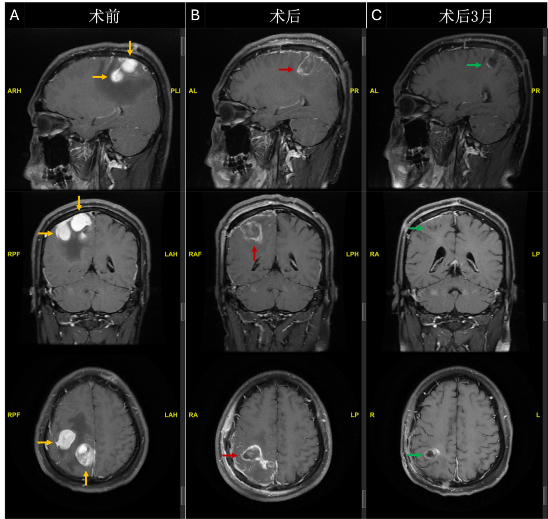
Serial brain MRI timeline. (A) Preoperative MRI showing multiple right parietal tumors (yellow arrows); (B) 1-week postoperative MRI showing surgical cavity with lace-like enhancement (red arrows); (C) 3-month follow-up MRI showing reduced enhancement and edema (green arrows), with no evidence of tumor recurrence.
Follow-Up Strategy
The treatment plan continues with the original 8-cycle ICE chemotherapy protocol. Follow-up monitoring includes:
- Contrast-enhanced brain MRI + SWI every 3 months post-surgery
- Regular assessment of tumor recurrence and neurological function
- Routine monitoring of blood counts, liver function, and renal function
Four Key Clinical Insights
- Rare CNS tumors require "morphology + molecular pathology" dual verification for diagnosis. For atypical intracranial mesenchymal tumors in young adults — especially those with spindle cell predominance, intratumoral hemorrhage, or dural tail signs — primary intracranial sarcoma must be strongly considered. NGS and DNA methylation profiling are the gold standard for definitive diagnosis and molecular classification.
- The MDT model is the optimal decision-making pathway for rare tumors without standard guidelines. This case successfully integrated surgical GTR evidence (HR = 0.061, 2-year OS 66.3%), molecular pathology-based exclusion of ineffective therapies (TMZ, MEKi), ICE regimen evidence from similar pediatric cases, and individualized assessment of blood-brain barrier permeability for chemotherapy accessibility.
- Comprehensive toxicity management is the safety guarantee for intensive sequential therapy. Intracranial sarcoma treatment is highly intensive. Standard chemoradiation can cause acute and long-term toxicities (myelosuppression, infection risk, hepatic/renal impairment, cardiac toxicity, secondary malignancies). Proactive planning and timely intervention — hepatic protection, prophylactic G-CSF, and early anti-infective management — ensured 5 cycles of ICE chemotherapy were completed without treatment interruption. This demonstrates that toxicity management is the fundamental prerequisite for delivering high-intensity comprehensive treatment.
- Many questions remain unanswered. The optimal number of chemotherapy cycles for adult intracranial sarcoma remains undefined (literature ranges from 2 to 8 cycles). The optimal sequencing of radiation and chemotherapy lacks comparative data. As global case registry data grows, multidisciplinary treatment guidelines for this entity may gradually emerge — particularly for adult patients, where balancing survival extension with cognitive and functional preservation will be a critical future research direction.
Expert Commentary: Prof. Zhang Junting, Beijing Tiantan Hospital
"This case report demonstrates a successful multidisciplinary team approach, combining maximal safe surgical resection with ICE chemotherapy and concurrent radiotherapy, in managing an extremely rare adult primary intracranial sarcoma (DICER1-mutant). In the short term, this achieved good tumor control and neurological functional preservation. This process deeply reflects the decisive role of molecular pathology in precision diagnosis and therapeutic exclusion, as well as the effectiveness of structured MDT processes in developing individualized strategies for rare tumors lacking standard guidelines."
"The primary diagnostic trap is that primary intracranial sarcoma (DICER1-mutant) often presents on imaging as a hypervascular mass with intratumoral hemorrhage — imaging features that overlap with CNS lymphoma or high-grade glioma. When biopsy specimens are limited, its diverse mesenchymal differentiation morphology can easily lead to misdiagnosis. Therefore, for young adults presenting with such atypical intracranial masses, molecular pathological diagnosis must be the core of the diagnostic workup."
"This case, through next-generation sequencing (NGS), confirmed biallelic DICER1 inactivation and TP53 co-mutation, achieving a gold-standard diagnosis. This emphasizes the indispensability of integrating morphology and molecular pathology for the precise diagnosis of rare intracranial mesenchymal tumors."
"From the treatment decision paradigm, this case successfully practiced the core value of MDT in managing rare tumors without standard guidelines. The MDT not only integrated evidence supporting maximal safe resection (GTR) as the strongest prognostic factor, but more critically, based on molecular pathology results, performed precise therapeutic exclusion and selection. For example, the unmethylated MGMT promoter status excluded temozolomide, while the wild-type RAS/RAF/MAPK pathway excluded MEK inhibitors."
"The final ICE chemotherapy regimen was based on efficacy observed in pediatric cases, and in this adult case was successfully implemented through proactive toxicity management (prophylactic G-CSF, hepatic protection). This highlights the importance of a comprehensive management strategy combining molecularly-guided systemic therapy with forward-looking supportive care."
"However, as a single-case report, the generalizability of these conclusions is limited. Follow-up time is relatively short, and long-term survival outcomes, late toxicities, and cognitive function impacts remain unknown. Furthermore, the optimal number of chemotherapy cycles for this entity in adults, and the ideal timing of radiation versus chemotherapy, remain unanswered. Future international registry and collaborative studies are urgently needed to accumulate more adult case data, enabling evidence-based treatment guidelines, with the core goal of optimizing survival while maximizing quality of life."
— Prof. Zhang Junting, Beijing Tiantan Hospital, Capital Medical University Affiliated
About the Case Breakthrough Notes Series
"Case Breakthrough Notes" (破局手记) is Arion Cancer Hospital's flagship clinical case series, documenting complex cancer cases where multidisciplinary collaboration achieved breakthroughs beyond standard treatment protocols. The series is guided by:
- Prof. Sun Min — Honorary Editor-in-Chief, UPMC Hillman Cancer Center
- Prof. Ma Zhiqiang — Honorary Editor-in-Chief, Arion Cancer Hospital GI Oncology Center
- Prof. Bai Li — Honorary Editor-in-Chief, Senior Oncology Expert
Column Editor: Prof. Gao Xiaofang
Contributing Author: Prof. Hong Ganlin
Expert Commentary: Prof. Zhang Junting, Beijing Tiantan Hospital (Capital Medical University)
References
- Louis DN, Perry A, Wesseling P, et al. The 2021 WHO Classification of Tumors of the Central Nervous System: a summary. Neuro Oncol. 2021;23(8):1231-1251.
- Vuong HG, Le MK, Dunn IF. A systematic review of the clinicopathological features and prognostic outcomes of DICER1-mutant malignant brain neoplasms. J Neurosurg Pediatr. 2022;30(3):308-315.
- Diaz Coronado RY, Mynarek M, Koelsche C, et al. Primary central nervous system sarcoma with DICER1 mutation — treatment results of a novel molecular entity in pediatric Peruvian patients. Cancer. 2022;128(4):697-707.
- Cardona AF, et al. DICER1-associated central nervous system sarcoma: A comprehensive clinical and genomic characterization of case series of young adult patients. Neurooncol Pract. 2023;10(4):381-390.
- Kosteniuk SE, Michaiel G, Dunham C. A Case of Primary Intracranial Sarcoma, DICER1-Mutant, in a Child with a Germline DICER1 Mutation. Brain Sci. 2023;13(7):1040.
- Lachance A, et al. Recurrent primary intracranial sarcoma, DICER1-mutant in a pediatric patient with DICER1 syndrome: the importance of molecular testing. Childs Nerv Syst. 2024;40(6):1965-1969.
- Koelsche C, et al. Primary intracranial spindle cell sarcoma with rhabdomyosarcoma-like features share a highly distinct methylation profile and DICER1 mutations. Acta Neuropathol. 2018;136(2):327-337.
- Kommoss FKF, et al. Genomic characterization of DICER1-associated neoplasms uncovers molecular classes. Nat Commun. 2023;14(1):1677.
- Honma H, et al. Primary intracranial sarcoma associated with DICER1 mutant: a case report and preclinical investigation. Brain Tumor Pathol. 2025;42(1):12-20.
- Zhang L, et al. RAS pathway targeted therapy in patients with DICER1-associated sarcomas. NPJ Precis Oncol. 2025;9(1):232.
Explore Advanced Neuro-Oncology Treatment at Arion
Our neuro-oncology MDT center, in collaboration with Beijing Tiantan Hospital, integrates neurosurgery, radiation oncology, molecular pathology, and medical oncology to deliver comprehensive care for the most challenging brain tumors — including rare and newly defined entities.